
Page 40 - Veterinary Histology of Domestic Mammals and Birds, 5th Edition
P. 40
22 Veterinary Histology of Domestic Mammals and Birds
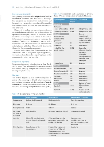
Endogenous pigments Table 1.2 Composition and occurrence of protein
VetBooks.ir Endogenous pigments are coloured products of cellular Type of protein Molecular Occurrence
classes (I–V) associated with intermediate filaments.
metabolism. In animal cells, these include haemoglo-
bin, myoglobin and iron reservoirs such as ferritin and
haemosiderin. Haemosiderin, a product of haemoglobin Class I & II weight
degradation, is stored in splenic macrophages or Kupffer
cells in the liver. Keratins:
Melanin is an endogenous pigment present in skin, – acid cytokeratins 40–64 All epithelial cells
the retinal pigment epithelium and in the meninges. In – basic cytokeratins 52–68 All epithelial cells
epidermal melanocytes, melanin is contained within Class III Cells of
membrane-bound organelles termed melanosomes. Vimentin and mesodermal
Differentiated melanosomes contain tyrosinase for vimentin-like origin (including
synthesis of melanin. As a dark pigment, melanin is pho- proteins: endothelial cells,
toprotective. The role of melanin in the function of the – vimentin 55 myofibroblasts)
retinal pigment epithelium (Figure 1.32) is described in – desmin 53 Muscle cells
Chapter 16, ‘Receptors and sense organs’. – glial fibrillary 50–52 Neuroglia
Residual bodies arising from fat metabolism are also acidic protein (oligodendroglia,
considered a form of endogenous pigment (lipofuscin). astrocytes,
Lipofuscin is often observed as a sign of ageing in cardiac microglia,
myocytes and in kidney and liver cells. ependymal cells),
Schwann cells,
pituicytes
Exogenous pigments
Exogenous pigments are primarily taken up from the air – peripherin 54 Neurons
via the lungs. They subsequently become concentrated Class IV
within tissue cells (e.g. soot particles or heavy metals in Neurofilament (L) 68 Neurons
the lungs or lymph nodes). Neurofilament (M) 110 Neurons
Neurofilament (H) 130 Neurons
Nucleus Synemin 182 Muscle cells
The nucleus (Figure 1.6) is an essential component of Paranemin 178 Muscle cells
animal cells, occurring in all cells other than mature Nestin 240 Muscle cells
mammalian erythrocytes. It is the repository of genetic Class V Nucleus of most
information, which is stored in coded form in chro- differentiated
mosomes containing deoxyribonucleic acid (DNA). Lamin A 62–72 cells
Lamin B 65–68 Nucleus of all
nucleated cells
Table 1.1 Characteristics of the cytoskeleton.
Actin filaments Microtubules Intermediate filaments
Appearance Helical double strand Hollow cylinder Cord-like bundles
Diameter 6–8 nm 20–25 nm 8–10 nm
Main protein(s) Actin Tubulin Various
Structural Thin, flexible Dynamic, transient (labile) Permanent (stable)
features of
filaments
Location Microvilli, terminal web, Cilia, centriole, spindle Desmosomes,
contractile units of muscle apparatus, contractile ring hemidesmosomes, in
cells during cell division association with nuclear
envelope
Main function Key element of the Intracellular transport, Structural
cytoskeleton movement of cilia, cell
shape and movement,
attachment of chromosomes
to spindle
Vet Histology.indb 22 16/07/2019 14:53