
Page 35 - Veterinary Histology of Domestic Mammals and Birds, 5th Edition
P. 35
The cell (cellula) 17
more flexible and shorter than microtubules. They consist · anchoring (and regulation of mobility) of membrane
VetBooks.ir of two forms of actin: free G-actin (globular actin) and proteins – actin filaments form a three-dimensional
polymerised F-actin (filamentous actin).
network throughout the cell and contribute to spe-
Polymerisation occurs at the rapidly growing plus end
+
2+
of the filament. This process requires K , Mg and ATP cialised lateral intercellular adhesions and other
types of anchoring junctions (e.g. focal adhesions),
and is regulated by various actin-binding proteins (ABP) · formation of the structural framework of microvilli,
(see below). · formation of the ‘terminal web’ beneath the cell
Actin filaments form a dense network that interconnects surface,
individual organelles. The filaments extend into periph- · cellular locomotion (e.g. migrating cells and tumour
eral cell processes and provide structural support for the cells) and
cytoplasm. In addition, they act together with myosin (tro- · formation of cell processes (e.g. filopodia) and con-
pomyosin) filaments to bring about cell contraction and tribution to cytoplasmic streaming.
associated motility. The specialised actin–myosin complex
of muscle cells is described in more detail in Chapter 4,
‘Muscle tissue’. Based on their diameter (5–7 nm), actin Microtubules
filaments are also referred to as microfilaments. Microtubules (Figures 1.26 and 1.30) are impermanent
Actin filaments undergo constant reorganisation accord- structures that can be rapidly assembled and dismantled.
ing to the functional demands of the cell. A number of Arising from the microtubule-organising centre (MTOC)
actin-binding proteins participate in this process (see also (containing the centrioles), microtubules are formed by
text box below). Cross-linking of actin filaments into paral- end-to-end attachment of free tubulin molecules. These
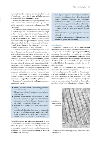
lel bundles by the actin-bundling protein fimbrin gives rise molecules originate from the disassembly of microtubules
to the structural core of specialised cell surface projections elsewhere in the cell. Microtubules also grow outward
known as microvilli and stereocilia (Figures 1.24 and 1.25). from basal bodies, the organising centre for cilia and fla-
Aggregates of actin filaments contribute to the contractile gella (see below).
ring that divides the cell during the final phase of mitosis. Microtubules are not contractile, rather they serve as
Actin filaments mediate processes associated with endo- and attachment sites for contractile proteins.
exocytosis, facilitate intramembranous movement of trans- Structurally, microtubules manifest as elongated, nar-
port proteins and expedite cellular movement. By combining row protein cylinders with a consistent diameter of 25
with filamin and α-actinin to form a flexible mesh, actin also nm. Measuring up to several micrometres in length, they
contributes to the gel-like nature of the cytoplasm. extend throughout the cell in an organised, cell-specific
Examples of non-muscle actin-binding proteins can be manner, thus contributing substantially to the morphol-
summarised as follows: ogy of the cell. When a cell undergoes a change in shape,
· fimbrin, villin and fascin: actin-bundling proteins as
seen in microvilli,
· filamin: cross-links with actin giving rise to gel-state
of cytoplasm,
· gelsolin: usually initiates polymerisation of actin,
but in the presence of high Ca concentrations
2+
causes severing of actin filaments, thus converting
the gel-like cytoplasm into a fluid state,
· vinculin: binds actin filaments to the plasmalemma,
and
· spectrin, ankyrin, adductin, protein 4.1 and protein
4.9: actin cross-linking proteins, studied particularly
in the cytoskeleton of erythrocytes, in which they
contribute to the stability of the cell membrane.
Actin filaments are not inherently contractile. It is the
interaction with numerous actin-binding proteins that
alters the spatial conformation of the filaments. The
proteins give the actin filaments specific characteristics,
enabling them to participate in a number of functions 1.24 Microvilli covered in glycocalyx (longitudinal sec-
within the cell, including: tion; x40,000).
Vet Histology.indb 17 16/07/2019 14:53