
Page 95 - Veterinary Histology of Domestic Mammals and Birds, 5th Edition
P. 95
Connective and supportive tissues (textus connectivus) 77
facilitate the intra-chondral transport of substances Chondrocytes are enclosed by a dense fibrillar colla-
VetBooks.ir involved in metabolism. gen network, giving rise to a thin region (1–2 μm) termed
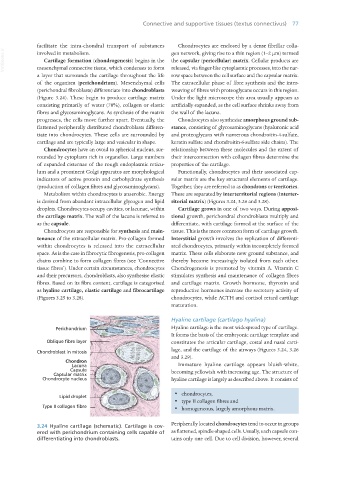
Cartilage formation (chondrogenesis) begins in the the capsular (pericellular) matrix. Cellular products are
mesenchymal connective tissue, which condenses to form released, via finger-like cytoplasmic processes, into the nar-
a layer that surrounds the cartilage throughout the life row space between the cell surface and the capsular matrix.
of the organism (perichondrium). Mesenchymal cells The extracellular phase of fibre synthesis and the inter-
(perichondral fibroblasts) differentiate into chondroblasts weaving of fibres with proteoglycans occurs in this region.
(Figure 3.24). These begin to produce cartilage matrix Under the light microscope this area usually appears as
consisting primarily of water (70%), collagen or elastic artificially expanded, as the cell surface shrinks away from
fibres and glycosaminoglycans. As synthesis of the matrix the wall of the lacuna.
progresses, the cells move further apart. Eventually, the Chondrocytes also synthesise amorphous ground sub-
flattened peripherally distributed chondroblasts differen- stance, consisting of glycosaminoglycans (hyaluronic acid
tiate into chondrocytes. These cells are surrounded by and proteoglycans with numerous chondroitin-4-sulfate,
cartilage and are typically large and vesicular in shape. keratin sulfate and chondroitin-6-sulfate side chains). The
Chondrocytes have an ovoid to spherical nucleus, sur- relationship between these molecules and the extent of
rounded by cytoplasm rich in organelles. Large numbers their interconnection with collagen fibres determine the
of expanded cisternae of the rough endoplasmic reticu- properties of the cartilage.
lum and a prominent Golgi apparatus are morphological Functionally, chondrocytes and their associated cap-
indicators of active protein and carbohydrate synthesis sular matrix are the key structural elements of cartilage.
(production of collagen fibres and glycosaminoglycans). Together, they are referred to as chondrons or territories.
Metabolism within chondrocytes is anaerobic. Energy These are separated by interterritorial regions (interter-
is derived from abundant intracellular glycogen and lipid ritorial matrix) (Figures 3.24, 3.26 and 3.28).
droplets. Chondrocytes occupy cavities, or lacunae, within Cartilage grows in one of two ways. During apposi-
the cartilage matrix. The wall of the lacuna is referred to tional growth, perichondral chondroblasts multiply and
as the capsule. differentiate, with cartilage formed at the surface of the
Chondrocytes are responsible for synthesis and main- tissue. This is the more common form of cartilage growth.
tenance of the extracellular matrix. Pro-collagen formed Interstitial growth involves the replication of differenti-
within chondrocytes is released into the extracellular ated chondrocytes, primarily within incompletely formed
space. As is the case in fibrocytic fibrogenesis, pro-collagen matrix. These cells elaborate new ground substance, and
chains combine to form collagen fibres (see ‘Connective thereby become increasingly isolated from each other.
tissue fibres’). Under certain circumstances, chondrocytes Chondrogenesis is promoted by vitamin A. Vitamin C
and their precursors, chondroblasts, also synthesise elastic stimulates synthesis and maintenance of collagen fibres
fibres. Based on its fibre content, cartilage is categorised and cartilage matrix. Growth hormone, thyroxin and
as hyaline cartilage, elastic cartilage and fibrocartilage reproductive hormones increase the secretory activity of
(Figures 3.25 to 3.28). chondrocytes, while ACTH and cortisol retard cartilage
maturation.
Hyaline cartilage (cartilago hyalina)
Hyaline cartilage is the most widespread type of cartilage.
It forms the basis of the embryonic cartilage template and
constitutes the articular cartilage, costal and nasal carti-
lage, and the cartilage of the airways (Figures 3.24, 3.26
and 3.29).
Immature hyaline cartilage appears bluish-white,
becoming yellowish with increasing age. The structure of
hyaline cartilage is largely as described above. It consists of:
· chondrocytes,
· type II collagen fibres and
· homogeneous, largely amorphous matrix.
3.24 Hyaline cartilage (schematic). Cartilage is cov- Peripherally located chondrocytes tend to occur in groups
ered with perichondrium containing cells capable of as flattened, spindle-shaped cells. Usually, each capsule con-
differentiating into chondroblasts. tains only one cell. Due to cell division, however, several
Vet Histology.indb 77 16/07/2019 14:56