Page 154 - Manual of Equine Field Surgery
        P. 154
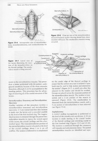
     150                        HEAD  AND NECK SURGERIES
                                              Cut  ends of:
                                          Ornohyoideus-ed
                                    Sternothyroideus
                                                                                                                                                       Omohyoideus  m.
                             Sternohyoideus  mm.
                                                                                                                                                   Figure 25-8  Close-up  view of the sternothyroideus
                                                                                                                                                   musculotendinous  section  showing  dotted  lines where
                                                                                                  -c~t;.;:i;~                                       resection will occur. The  omohyoideus muscle is being
                        Figure 25-6  Intraoperative  view  of  sternohyoidec-                                                                      retracted.
                        tomy,  sternothyroidectomy,  and  omohyoidectomy  for
                        DDSP.
                                                                                                                                                      Epiglottis              Arytenoid  cartilage
                                                                                                               Ceratohyoideus  m.
                                                                                                                                                                                                                       Cricoid  cartilage
                                                                                                                       Ceratohyoid
                         Figure  25- 7  Lateral  view  of                                             Lingual  process               bone
                         the  larynx  illustrating  the  inser-                                   of basihyoid  bone
                         tion  of  the  sternothyroideus  on                                                                        ---~.,,....,:;
                         the  thyroid  cartilage.  The  omohy-                                                                Thyrohyoid
                         oideus  muscle is being retracted.                                                                               bone  Thyrohyoid
                                                                                                                                                      membrane                                                        Retraction  of
                                                                                                                                                                         Insertion  of                 the omohyoideus  m.
                                                                                                                                                                sternothyroideus  m.
                         ments  to  the sternohyoideus  muscles. This proce-                                                                         on  the  caudal  edge  of  the  thyroid  cartilage  to
                          dure  is better  performed  with  the  patient  under                                                                      ensure that the correct  structure  is isolated.  A spay
                          general  anesthesia  because  of the  more  extensive                                                                      hook  may be  used  to help  isolate  and  exteriorize
                          dissection,  although  it can be accomplished  in the                                                                      the  tendon9  (Figure 25-7).  A small vein  often  lies
                          standing  patient.  This  procedure  has  the  advan-                                                                      adjacent  to  the  tendon  and  should  be  avoided.
                          tage  of removing  all of the caudal retractors of the                                                                     Forceps are placed  across the muscular  portion of
                          larynx.                                                                                                                    the  sternothyroideus  muscle.  The tendon  is tran-
                                                                                                                                                     sected,  and  a  2-cm  portion  of  the  muscle  is
                          Sternothyroideus  Tenectomy and Sternohyoideus                                                                             removed  (Figure  25-8).                                     The  omohyoideus  is
                          Myectomy                                                                                                                    dissected  from  the  sternohyoideus  muscle,  and  a
                          Another  variation  of  this  procedure  involves  a                                                                        5-cm  section  of sternohyoideus  is  then  removed
                          sternothyroideus  tenectomy  and  sternohyoideus                                                                            (see Figure 25-6).
                          myectomy,  A 5-cm  ventral mid-line  skin  incision
                           is made over the  larynx  and  is extended  caudally                                                                       Staphylectomy
                           to the  level of the  first tracheal  ring. The longitu-                                                                   A  ventral  midline  laryngotomy  is  performed  at
                           dinal incision  is extended  through the paired ster-                                                                      the  level of the cricothyroid  membrane.  A 10-cm
                           nohyoideus  muscles  to  expose  the  ventral  aspect                                                                      incision  is  made  starting  at  the  cranial  border
                           of the  larynx, the  cricoid  cartilage,  and  the  crico-                                                                 of  the  thyroid  cartilage  and  extending  caudal  to
                           tracheal  space. The  musculotendinous  portion  of                                                                        the  first  tracheal  ring  (Figures  25-9  and  25-10).
                           the sternothyroideus  muscle is located  at the level                                                                      The incision  is continued  through  the  cutaneous
                           of  the  cricoid  cartilage,  about  3  to  4  cm  off                                                                     colli muscles  and  subcutaneous  tissue. The paired
                           midline.  The  tendon is followed to its attachment                                                                        sternohyoideus  muscles  are  identified  and  sepa-


