Page 245 - Manual of Equine Field Surgery
P. 245

Rectovagina Fistula  Repair                                         241
                                                                                                                                                                                                          l


                       Sutures  are  positioned  approximately  1  cm apart,


                       avoiding  the vaginal  and  rectal  mucosa.  Once  the


                       cranial  half  of  the  rectovaginal  shelf  is  recon-


                       structed,  closure  of  the  vaginal  mucosa  is  corn-


                       pleted,  followed  by  closure  of  the  remaining


                       caudal  half  of the  rectovaginal  shelf.  The  trans-


                       verse perinea!  skin incision  is closed  with  No. 2-0


                       nonabsorbable  suture  using  a simple interrupted


                       pattern.  If indicated,  Caslick's  procedure  is  per-


                       formed to appose the vulvar opening.


                       Klug Technique


                       Fistulas  up to  6  cm have been repaired  using  this


                                              7
                      technique.  The  Klug  technique  provides  good

                      visualization,  a  durable  and  stable  closure,  and


                       good  first-time  healing  success  rate  without  dis-


                       rupting the integrity of the anal sphincter.  Repair-


                      ing  a large  fistula  using this  technique  should  be


                       attempted  with  caution.  Tissue  mobilization  is                                                                                 A


                       difficult in large fistula  repairs  and may require an                                                                                               -


                       alternative repair method.  Cranially located  fistu-


                      las  should  not  be  repaired  with  this  technique                                                                                                   ·=::_  )


                      because  of  inadequate  visualization  and  limited


                      working room.                                                                                                                                          B


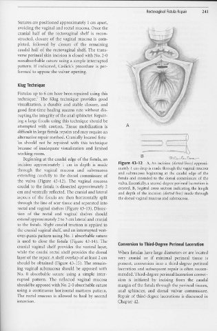
                             Beginning  at the  caudal edge of the  fistula,  an


                      incision  approximately  1  cm  in  depth  is  made                                                                         Figure 43-12                      A, An  incision  ( dotted lines) approxi-


                      through  the  vaginal  mucosa  and  subrnucosa                                                                              mately 1  cm deep is made through  the vaginal mucosa
                                                                                                                                                  and  submucosa  beginning  at the  caudal  edge  of the
                      extending  caudally  to  the  dorsal  cornmissure  of                                                                       fistula and extended  to  the dorsal  commissure  of the


                      the  vulva  (Figure  43-12).  The  vaginal  mucosa                                                                         vulva.  Essentially, a second-degree perineal laceration is


                      caudal  to  the fistula  is  dissected  approximately  2                                                                    created.  B,  Sagittal  cross  section  indicating  the  length


                      cm and ventrally reflected.  The  cranial and  lateral                                                                      and  depth  of the  incision  (dotted line)  made  through


                      aspects  of the  fistula  are  then  horizontally  split                                                                    the dorsal vaginal mucosa and submucosa.


                      through  the  line of scar tissue  and  separated  into


                      rectal and vaginal  shelves  (Figure  43-13  ).  Dissec-


                      tion  of  the  rectal  and  vaginal  shelves  should


                      extend approximately 2 to 3 cm lateral and cranial


                      to the fistula.  Slight  caudal traction  is  applied  to


                      the cranial  vaginal  shelf, and  an interrupted  vest-


                      over-pants  pattern  using  No.  1  absorbable  suture


                      is  used  to  close  the  fistula  (Figure  43-14).  The                                                                   Conversion to Third-Degree  Perineal  Laceration


                      cranial  vaginal  shelf  provides  the  ventral  layer,


                      while  the  caudal  rectal  shelf  provides  the  dorsal                                                                   When  fistulas  have large diameters  or  are  located


                      layer of the repair.  A shelf overlap of at least  2 cm                                                                    very  cranial  or  if  minimal  perineal  tissue  is


                      should  be  obtained  (Figure  43-15).  The  remain-                                                                       present,  conversion  into  a  third-degree  perinea!


                      ing  vaginal  submucosa  should  be  apposed  with                                                                         laceration  and  subsequent  repair  is often  recom-


                      No.  0  absorbable  suture  11siI1g  a  simple  inter-                                                                     mended.Third-degree  perineal  laceration  conver-


                      rupted  pattern.  The  reflected  vaginal  mucosa                                                                          sion  is  initiated  by  incising  from  the  caudal


                      should  be apposed  with No. 2-0  absorbable  suture                                                                       margin  of the fistula through  the perineal  tissues,


                      using  a  continuous  horizontal  mattress  pattern.                                                                       anal  sphincter,  and  dorsal  vulvar  commissure.


                      The  rectal  mucosa  is  allowed  to  heal  by  second                                                                     Repair  of third-degree  lacerations  is discussed  in


                      intention.                                                                                                                 Chapter  42.
   240   241   242   243   244   245   246   247   248   249   250