
Page 1049 - Clinical Small Animal Internal Medicine
P. 1049
109 Fungal Infections 987
reversed and aggressive antifungal drug treatment is
VetBooks.ir initiated promptly, complete recovery is possible.
Public Health Implications
Direct inoculation of yeast forms of fungi can potentially
cause infection. The mycelial form of dimorphic fungi
can pose significant zoonotic risk, although these are
typically only present when the organisms are cultured
in the laboratory. Appropriate precautions such as avoid
ing needle sticks and other traumatic wounds when
working with animals that have deep fungal infections
should be followed. Caution when performing necrop
sies, and cremating, rather than allowing burial of
deceased patients are also important to prevent zoonosis
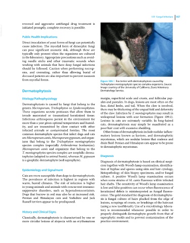
from mycelial forms. Figure 109.1 Fox terrier with dermatophytosis caused by
Trichophyton mentagrophytes species complex organisms. Source:
Image courtesy of the University of California, Davis Veterinary
Dermatophytosis Dermatology Service.
Etiology/Pathophysiology margin, superficial scale and crusts, and follicular pap
ules and pustules. In dogs, lesions are most often on the
Dermatophytosis is caused by fungi that belong to the face, distal limbs, and tail. When the claw is involved,
genera Microsporum, Trichophyton or Epidermophyton. there may be thickening of the ungual fold and deformity
These organisms secrete proteases that allow them to of the claw. Infection by T. mentagrophytes may result in
invade macerated or traumatized keratinized tissue. widespread lesions with scar formation (Figure 109.1).
Infectious arthrospores persist in the environment for Lesions in cats are extremely variable. In long‐haired
more than a year given optimal temperature and humid cats, dermatophytosis may simply be manifested as a
ity, and are transmitted by close contact with other poor hair coat with excessive shedding.
infected animals or contaminated fomites. The most Other forms of dermatophytosis include nodular inflam
common dermatophyte species that infect dogs and cats matory lesions known as kerions, and dermatophytic
are Microsporum canis, Microsporum gypseum, and organ mycetomas, which are nodular lesions that coalesce and
isms that belong to the Trichophyton mentagrophytes drain fluid. Persian and Himalayan cats appear to be prone
species complex (especially Arthroderma benhamiae). to dermatophytic mycetomas.
Microsporum canis and organisms that belong to the
T. mentagrophytes species complex are zoophilic derma
tophytes (adapted to animal hosts), whereas M. gypseum Diagnosis
is a geophilic dermatophyte (soil saprophyte). Diagnosis of dermatophytosis is based on clinical suspi
cion together with Wood’s lamp examination, identifica
Epidemiology and Signalment tion of hyphae and spores using skin and hair cytology,
histopathology of skin biopsy specimens, and/or fungal
Cats are more susceptible than dogs to dermatophytosis. culture. A positive Wood’s lamp examination occurs
The prevalence of infection is highest in regions with when some strains of M. canis fluoresce within infected
warm, humid climates. The risk of infection is higher hair shafts. The sensitivity of Wood’s lamp examination
in young animals and animals with concurrent immuno is low and false positives can occur when fluorescence of
suppressive disorders, such as hyperadrenocorticism. keratinized debris is misinterpreted as fungal fluores
Dogs that burrow in soil may also be at increased risk. cence. The gold standard for diagnosis of dermatophyto
Persian and Himalayan cats and Yorkshire and Jack sis is fungal culture of hairs plucked from the edge of
Russell terriers appear to be predisposed. lesions, scrapings of crusts, or brushings of the haircoat
(using a new toothbrush). Use of a microbiology labora
tory is recommended whenever possible in order to
History and Clinical Signs
properly distinguish dermatophyte growth from that of
Classically, dermatophytosis is characterized by one or saprophytic molds and to prevent contamination of the
more circular lesions of alopecia with an erythematous practice environment.