
Page 1052 - Clinical Small Animal Internal Medicine
P. 1052
990 Section 9 Infectious Disease
alveolar‐interstitial patterns, pulmonary mass lesions, involvement, radiographic lesions and clinical signs may
VetBooks.ir tracheobronchial lymphadenopathy, and/or single or initially worsen with treatment. Initial worsening of radi
ographic lesions does not appear to affect survival. Urine
multiple pulmonary nodules that resemble pulmonary
neoplasia. Radiographs of affected bone typically show
Although a positive residual urine antigen titer at the
osteolytic lesions, often accompanied by periosteal antigen concentration declines with effective treatment.
proliferation and soft tissue swelling. time treatment is discontinued does not necessarily pre
A diagnosis of blastomycosis is often confirmed dict that relapse will occur, a negative urine antigen test
through cytologic examination of affected tissues, lung is recommended before treatment is discontinued. Most
washes or body fluids. The yeasts are 8–15 μm in diameter, dogs require at least 4–6 months of treatment, and some
have a thick, refractile cell wall, and exhibit broad‐based dogs may require treatment for more than one year,
budding, although occasionally organisms are not visual especially those with osteoarticular infections or wide
ized. The organisms are usually accompanied by pyo spread dissemination. Treatment with amphotericin B
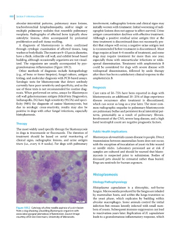
granulomatous inflammation (Figure 109.2). could be considered for dogs with severe disease with
Other methods of diagnosis include histopathology widespread dissemination, followed by azole therapy
(e.g., of bone or tissue biopsies), fungal culture, antigen after there has been a satisfactory clinical response to the
testing, and molecular diagnosis with PCR‐based assays. amphotericin B.
Serologic tests for blastomycosis that detect antibody
currently have poor sensitivity and specificity, and so the
use of these tests is not recommended for routine diag Prognosis
nosis. When performed on urine, assays for Blastomyces Cure rates of 50–75% have been reported in dogs with
cell wall galactomannan antigen (MiraVista Diagnostics, blastomycosis; an additional 20–25% of dogs experience
Indianapolis, IN) have high sensitivity (93.5%) and speci disease recurrence after treatment is discontinued,
ficity (98%) for diagnosis of canine blastomycosis, but which can occur as long as a year later. The most com
due to serologic cross‐reactivity, results may also be mon radiographic sequelae to pulmonary blastomycosis
positive in dogs with other fungal infections, especially are pulmonary bullae and persistent focal interstitial pat
histoplasmosis. terns, presumably as a result of pulmonary fibrosis.
Involvement of the CNS, severe lung disease, and a high
Therapy band neutrophil count are negative prognostic factors.
The most widely used specific therapy for blastomycosis
in dogs is itraconazole or fluconazole. The duration of Public Health Implications
treatment should be based on serial monitoring of Blastomyces dermatitidis causes disease in people. Direct
clinical signs, radiographic lesions, and urine antigen transmission between mammalian hosts does not occur,
titers (i.e., every 4–8 weeks). For dogs with pulmonary with the exception of inoculation of yeast via bite wound
or needle sticks. Laboratory personnel are at risk if
samples are cultured and should be warned that blasto
mycosis is suspected prior to submission. Bodies of
deceased pets should be cremated rather than buried.
Dogs are sentinels for human exposure.
Histoplasmosis
Etiology/Pathophysiology
Histoplasma capsulatum is a dimorphic, soil‐borne
fungus. Microconidia produced by the fungus are inhaled
by mammalian hosts, and within the lungs transition to
the yeast phase, which replicates by budding within
10 m alveolar macrophages. Some animals control the initial
infection but remain latently infected with small num
Figure 109.2 Cytology of a fine needle aspirate of a skin lesion bers of yeasts. Subsequent immune suppression can lead
from a dog showing a budding Blastomyces organism with
associated pyogranulomatous inflammation. Source: Image to reactivation years later. Replication of H. capsulatum
courtesy of Dr Jed Overmann, University of Minnesota. leads to a granulomatous inflammatory response, which