
Page 1053 - Clinical Small Animal Internal Medicine
P. 1053
109 Fungal Infections 991
is sometimes accompanied by fibrosis. When a cell‐ inflammation. Dogs with acute progressive disseminated
VetBooks.ir mediated immune response is defective, yeast forms histoplasmosis may have increased band neutrophils,
lymphopenia, monocytopenia, and thrombocytopenia.
migrate to local lymph nodes (such as the tracheobron
chial lymph nodes) and other tissues that contain mono
and evidence of coagulopathies may be present in ani
nuclear cells, such as the liver and spleen. Other common Increased liver enzyme activities, hyperbilirubinemia,
sites of dissemination include the bone marrow, small mals with hepatic involvement. In cats, plain thoracic
and/or large intestinal tract, pancreas, the skin, bones, radiographs can reveal diffuse, linear, nodular or miliary
central nervous system, and eyes. interstitial patterns. Alveolar, interstitial, and/or bron
chial patterns, tracheobronchial lymphadenopathy, lung
lobe consolidation, and/or rarely pleural effusion can be
Epidemiology and Signalment
seen in dogs. Lesions in dogs can sometimes calcify.
Histoplasma capsulatum is found worldwide, but espe Abdominal imaging may show hepatomegaly, spleno
cially along the Mississippi, Missouri, Tennessee, and megaly, abdominal lymphadenopathy or ascites. A thick
Ohio river valleys of the United States as well as in Latin ened intestinal wall with architectural disruption of the
America. H. capsulatum can be found in the intestinal bowel wall may be identified on ultrasound examination.
tract and guano of bats, which constitute the primary Colonoscopic findings in dogs with colonic histoplasmo
reservoir of the organism and disseminate it geographi sis include irregularity, ulceration, increased granularity,
cally. H. capsulatum can also be found in high concentra and friability of the colonic wall.
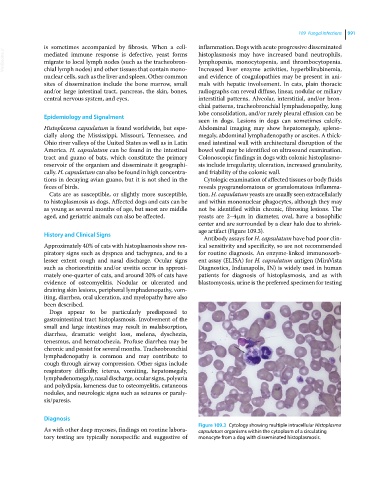
tions in decaying avian guano, but it is not shed in the Cytologic examination of affected tissues or body fluids
feces of birds. reveals pyogranulomatous or granulomatous inflamma
Cats are as susceptible, or slightly more susceptible, tion. H. capsulatum yeasts are usually seen extracellularly
to histoplasmosis as dogs. Affected dogs and cats can be and within mononuclear phagocytes, although they may
as young as several months of age, but most are middle not be identified within chronic, fibrosing lesions. The
aged, and geriatric animals can also be affected. yeasts are 2–4 μm in diameter, oval, have a basophilic
center and are surrounded by a clear halo due to shrink
age artifact (Figure 109.3).
History and Clinical Signs
Antibody assays for H. capsulatum have had poor clin
Approximately 40% of cats with histoplasmosis show res ical sensitivity and specificity, so are not recommended
piratory signs such as dyspnea and tachypnea, and to a for routine diagnosis. An enzyme‐linked immunosorb
lesser extent cough and nasal discharge. Ocular signs ent assay (ELISA) for H. capsulatum antigen (MiraVista
such as chorioretinitis and/or uveitis occur in approxi Diagnostics, Indianapolis, IN) is widely used in human
mately one‐quarter of cats, and around 20% of cats have patients for diagnosis of histoplasmosis, and as with
evidence of osteomyelitis. Nodular or ulcerated and blastomycosis, urine is the preferred specimen for testing
draining skin lesions, peripheral lymphadenopathy, vom
iting, diarrhea, oral ulceration, and myelopathy have also
been described.
Dogs appear to be particularly predisposed to
gastrointestinal tract histoplasmosis. Involvement of the
small and large intestines may result in malabsorption,
diarrhea, dramatic weight loss, melena, dyschezia,
tenesmus, and hematochezia. Profuse diarrhea may be
chronic and persist for several months. Tracheobronchial
lymphadenopathy is common and may contribute to
cough through airway compression. Other signs include
respiratory difficulty, icterus, vomiting, hepatomegaly,
lymphadenomegaly, nasal discharge, ocular signs, polyuria
and polydipsia, lameness due to osteomyelitis, cutaneous
nodules, and neurologic signs such as seizures or paraly
sis/paresis.
Diagnosis
Figure 109.3 Cytology showing multiple intracellular Histoplasma
As with other deep mycoses, findings on routine labora capsulatum organisms within the cytoplasm of a circulating
tory testing are typically nonspecific and suggestive of monocyte from a dog with disseminated histoplasmosis.