
Page 1137 - Clinical Small Animal Internal Medicine
P. 1137
118 Disorders of Sodium and Water Homeostasis 1075
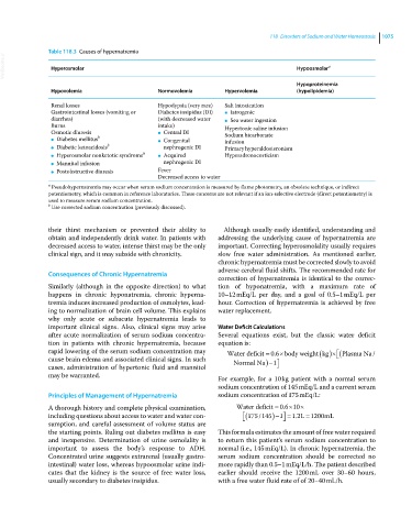
Table 118.3 Causes of hypernatremia
VetBooks.ir Hyperosmolar Hypoosmolar a
Hypoproteinemia
Hypovolemia Normovolemia Hypervolemia (hypolipidemia)
Renal losses Hypodypsia (very rare) Salt intoxication
Gastrointestinal losses (vomiting or Diabetes insipidus (DI) ● Iatrogenic
diarrhea) (with decreased water ● Sea water ingestion
Burns intake) Hypertonic saline infusion
Osmotic diuresis ● Central DI Sodium bicarbonate
● Diabetes mellitus b ● Congenital infusion
● Diabetic ketoacidosis b nephrogenic DI Primary hyperaldosteronism
● Hyperosmolar nonketotic syndrome b ● Acquired Hyperadrenocorticism
● Mannitol infusion nephrogenic DI
● Postobstructive diuresis Fever
Decreased access to water
a Pseudohypernatremia may occur when serum sodium concentration is measured by flame photometry, an obsolete technique, or indirect
potentiometry, which is common in reference laboratories. These concerns are not relevant if an ion‐selective electrode (direct potentiometry) is
used to measure serum sodium concentration.
b Use corrected sodium concentration (previously discussed).
their thirst mechanism or prevented their ability to Although usually easily identified, understanding and
obtain and independently drink water. In patients with addressing the underlying cause of hypernatremia are
decreased access to water, intense thirst may be the only important. Correcting hyperosmolality usually requires
clinical sign, and it may subside with chronicity. slow free water administration. As mentioned earlier,
chronic hypernatremia must be corrected slowly to avoid
adverse cerebral fluid shifts. The recommended rate for
Consequences of Chronic Hypernatremia
correction of hypernatremia is identical to the correc-
Similarly (although in the opposite direction) to what tion of hyponatremia, with a maximum rate of
happens in chronic hyponatremia, chronic hyperna- 10–12 mEq/L per day, and a goal of 0.5–1 mEq/L per
tremia induces increased production of osmolytes, lead- hour. Correction of hypernatremia is achieved by free
ing to normalization of brain cell volume. This explains water replacement.
why only acute or subacute hypernatremia leads to
important clinical signs. Also, clinical signs may arise Water Deficit Calculations
after acute normalization of serum sodium concentra- Several equations exist, but the classic water deficit
tion in patients with chronic hypernatremia, because equation is:
rapid lowering of the serum sodium concentration may Water deficit 06 body weightkg Plasma Na/
.
cause brain edema and associated clinical signs. In such Normal Na 1 1
cases, administration of hypertonic fluid and mannitol
may be warranted. For example, for a 10 kg patient with a normal serum
sodium concentration of 145 mEq/L and a current serum
Principles of Management of Hypernatremia sodium concentration of 175 mEq/L:
.
A thorough history and complete physical examination, Water deficit 06 10
including questions about access to water and water con- 175 145 1 1 2 L 1200 mL
/
.
sumption, and careful assessment of volume status are
the starting points. Ruling out diabetes mellitus is easy This formula estimates the amount of free water required
and inexpensive. Determination of urine osmolality is to return this patient’s serum sodium concentration to
important to assess the body’s response to ADH. normal (i.e., 145 mEq/L). In chronic hypernatremia, the
Concentrated urine suggests extrarenal (usually gastro- serum sodium concentration should be corrected no
intestinal) water loss, whereas hypoosmolar urine indi- more rapidly than 0.5–1 mEq/L/h. The patient described
cates that the kidney is the source of free water loss, earlier should receive the 1200 mL over 30–60 hours,
usually secondary to diabetes insipidus. with a free water fluid rate of of 20–40 mL/h.