
Page 1146 - Clinical Small Animal Internal Medicine
P. 1146
1084 Section 10 Renal and Genitourinary Disease
Studies suggest that up to 43% of cats with hyperthy
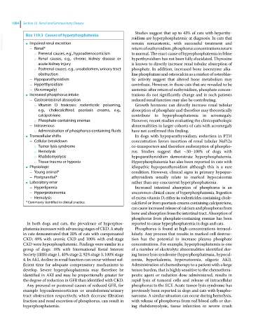
Box 119.3 Causes of hyperphosphatemia
VetBooks.ir ● Impaired renal excretion roidism are hyperphosphatemic at diagnosis. In cats that
remain nonazotemic, with successful treatment and
return of euthyroidism, phosphorus concentrations return
– Renal*
○ Prerenal causes, e.g., hypoadrenocorticism to normal. The exact cause of hyperphosphatemia in feline
○ Renal causes, e.g., chronic kidney disease or hyperthyroidism has not been fully elucidated. Thyroxine
acute kidney injury is known to directly increase renal tubular absorption of
○ Postrenal causes, e.g., uroabdomen, urinary tract phosphate. In addition, increased bone isoenzyme alka
obstruction line phosphatase and osteocalcin as a marker of osteoblas
– Hypoparathyroidism tic activity suggest that altered bone metabolism may
– Hyperthyroidism contribute. However, in those cats that are revealed to be
– (Acromegaly) azotemic after return of euthyroidism, phosphate concen
Increased phosphorus intake trations do not significantly change and in such patients
●
– Gastrointestinal absorption reduced renal function may also be contributing.
○ Vitamin D toxicosis: rodenticide poisoning, Growth hormone can directly increase renal tubular
e.g., cholecalciferol; psoriasis creams, e.g., absorption of phosphate and therefore may theoretically
calcipotriene contribute to hyperphosphatemia in acromegaly.
○ Phosphate‐containing enemas However, recent studies evaluating the clinicopathologic
– Intravenous abnormaltities in larger cohorts of cats with acromegaly
○ Administration of phosphorus‐containing fluids have not confirmed this finding.
Transcellular shifts In dogs with hypoparathyroidism, reduction in PTH
●
– Cellular breakdown concentration favors insertion of renal tubular NaPi2a
○ Tumor lysis syndrome co‐transporters and therefore reabsorption of phospho
○ Hemolysis rus. Studies suggest that ~30–100% of dogs with
○ Rhabdomyolysis hypoparathyroidism demonstrate hyperphosphatemia.
○ Tissue trauma or hypoxia Hyperphosphatemia has also been reported in cats with
Physiologic idiopathic hypoparathyroidism although this is a rare
●
– Young animal* condition. However, clinical signs in primary hypopar
– Postprandial* athyroidism usually relate to marked hypocalcemia
Laboratory error rather than any concurrent hyperphosphatemia.
●
– Hyperlipemia Increased intestinal absorption of phosphorus is an
– Hyperproteinemia uncommon clinical cause of hyperphosphatemia. Ingestion
– Hemolysis of excess vitamin D, either in rodenticides containing chole
* Commonly identified in clinical practice. calciferol or from psoriasis creams containing calcipotriene,
can cause increased release of calcium and phosphorus from
bone and absorption from the intestinal tract. Absorption of
phosphorus from phosphate‐containing enemas has been
In both dogs and cats, the prevalence of hyperphos reported to cause hyperphosphatemia in dogs and cats.
phatemia increases with advancing stages of CKD. A study Phosphorus is found at high concentrations intracel
in cats demonstrated that 20% of cats with compensated lularly. Any process that results in marked cell destruc
CKD, 49% with uremic CKD and 100% with end‐stage tion has the potential to increase plasma phosphate
CKD were hyperphosphatemic. Findings were similar in a concentrations. For example, hyperphosphatemia is one
group of dogs: 18% with International Renal Interest of a number of electrolyte abnormalities identified dur
Society (IRIS) stage 1, 40% stage 2, 92% stage 3, 100% stage ing tumor lysis syndrome (hyperphosphatemia, hypocal
4. In AKI, decline in renal function can occur without suf cemia, hyperkalemia, hyperuricemia, oliguric AKI).
ficient time for adequate compensatory mechanisms to Administration of chemotherapy to a patient with a large
develop. Severe hyperphosphatemia may therefore be tumor burden, that is highly sensitive to the chemothera
identified in AKI and may be proportionally greater for peutic agent or radiation dose administered, results in
the degree of reduction in GFR than identified with CKD. rapid lysis of tumoral cells and release of intracellular
Any prerenal or postrenal causes of reduced GFR, for phosphorus to the ECF. Acute tumor lysis syndrome has
example hypoadrenocorticism or uroabdomen/urinary previously been reported in dogs and cats with lympho
tract obstruction respectively, which decrease filtration sarcoma. A similar situation can occur during hemolysis,
fraction and renal excretion of phosphorus, can result in with release of phosphorus from red blood cells or dur
hyperphosphatemia. ing rhabdomyolysis, tissue infarction or severe crush