
Page 1144 - Clinical Small Animal Internal Medicine
P. 1144
1082 Section 10 Renal and Genitourinary Disease
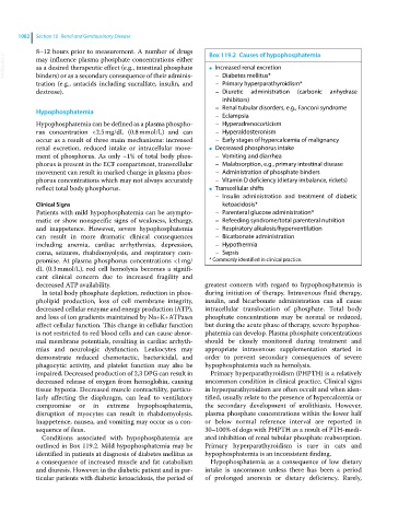
8–12 hours prior to measurement. A number of drugs Box 119.2 Causes of hypophosphatemia
VetBooks.ir may influence plasma phosphate concentrations either ● Increased renal excretion
as a desired therapeutic effect (e.g., intestinal phosphate
binders) or as a secondary consequence of their adminis
tration (e.g., antacids including sucralfate, insulin, and – Diabetes mellitus*
– Primary hyperparathyroidism*
dextrose). – Diuretic administration (carbonic anhydrase
inhibitors)
– Renal tubular disorders, e.g., Fanconi syndrome
Hypophosphatemia
– Eclampsia
Hypophosphatemia can be defined as a plasma phospho – Hyperadrenocorticism
rus concentration <2.5 mg/dL (0.8 mmol/L) and can – Hyperaldosteronism
occur as a result of three main mechanisms: increased – Early stages of hypercalcemia of malignancy
renal excretion, reduced intake or intracellular move ● Decreased phosphorus intake
ment of phosphorus. As only ~1% of total body phos – Vomiting and diarrhea
phorus is present in the ECF compartment, transcellular – Malabsorption, e.g., primary intestinal disease
movement can result in marked change in plasma phos – Administration of phosphate binders
phorus concentrations which may not always accurately – Vitamin D deficiency (dietary imbalance, rickets)
reflect total body phosphorus. ● Transcellular shifts
– Insulin administration and treatment of diabetic
Clinical Signs ketoacidosis*
Patients with mild hypophosphatemia can be asympto – Parenteral glucose administration*
matic or show nonspecific signs of weakness, lethargy, – Refeeding syndrome/total parenteral nutrition
and inappetence. However, severe hypophosphatemia – Respiratory alkalosis/hyperventilation
can result in more dramatic clinical consequences – Bicarbonate administration
including anemia, cardiac arrhythmias, depression, – Hypothermia
coma, seizures, rhabdomyolysis, and respiratory com – Sepsis
promise. At plasma phosphorus concentrations <1 mg/ * Commonly identified in clinical practice.
dL (0.3 mmol/L), red cell hemolysis becomes a signifi
cant clinical concern due to increased fragility and
decreased ATP availability. greatest concern with regard to hypophosphatemia is
In total body phosphate depletion, reduction in phos during initiation of therapy. Intravenous fluid therapy,
pholipid production, loss of cell membrane integrity, insulin, and bicarbonate administration can all cause
decreased cellular enzyme and energy production (ATP), intracellular translocation of phosphate. Total body
and loss of ion gradients maintained by Na+K+ATPases phosphate concentrations may be normal or reduced,
affect cellular function. This change in cellular function but during the acute phase of therapy, severe hypophos
is not restricted to red blood cells and can cause abnor phatemia can develop. Plasma phosphate concentrations
mal membrane potentials, resulting in cardiac arrhyth should be closely monitored during treatment and
mias and neurologic dysfunction. Leukocytes may appropriate intravenous supplementation started in
demonstrate reduced chemotactic, bactericidal, and order to prevent secondary consequences of severe
phagocytic activity, and platelet function may also be hypophosphatemia such as hemolysis.
impaired. Decreased production of 2,3 DPG can result in Primary hyperparathyroidism (PHPTH) is a relatively
decreased release of oxygen from hemoglobin, causing uncommon condition in clinical practice. Clinical signs
tissue hypoxia. Decreased muscle contractility, particu in hyperparathyroidism are often occult and when iden
larly affecting the diaphragm, can lead to ventilatory tified, usually relate to the presence of hypercalcemia or
compromise or in extreme hypophosphatemia, the secondary development of urolithiasis. However,
disruption of myocytes can result in rhabdomyolysis. plasma phosphate concentrations within the lower half
Inappetence, nausea, and vomiting may occur as a con or below normal reference interval are reported in
sequence of ileus. 30–100% of dogs with PHPTH as a result of PTH‐medi
Conditions associated with hypophosphatemia are ated inhibition of renal tubular phosphate reabsorption.
outlined in Box 119.2. Mild hypophosphatemia may be Primary hyperparathyroidism is rare in cats and
identified in patients at diagnosis of diabetes mellitus as hypophosphatemia is an inconsistent finding.
a consequence of increased muscle and fat catabolism Hypophosphatemia as a consequence of low dietary
and diuresis. However, in the diabetic patient and in par intake is uncommon unless there has been a period
ticular patients with diabetic ketoacidosis, the period of of prolonged anorexia or dietary deficiency. Rarely,