
Page 1183 - Clinical Small Animal Internal Medicine
P. 1183
122 Obstructive Uropathy 1121
right ureteral obstruction. In addition, it had a severely devices are commercially available. Subcutaneous ure
VetBooks.ir shrunken left kidney, which was likely nonfunctional. teral bypass employs a nephrostomy tube and a cystos
tomy tube, which are connected to a dual‐lumen vascular
After placement of the right ureteral stent, the cat was
discharged four days later with a BUN of 16 mg/dL and a
wall. This port allows the system to be flushed at regular
creatinine of 1.4 mg/dL. port that is attached under the skin to the ventral body
In the authors’ experience, ureteral stents in cats pro intervals to avoid obstruction and facilitates urine sam
vide acute resolution of the obstruction but have a mod pling (see Figure 122.11). At many centers, subcutaneous
erate risk for ureteral irritation and pollakiuria that can ureteral bypass devices are now the treatment of choice
be challenging to manage. Studies suggest one‐third of for obstructive urolithiasis in cats but are rarely implanted
cats with a ureteral stent will have dysuria either tran in dogs where conventional surgery or temporary ureteral
siently or permanently and that 19% of feline ureteral stenting are often effective. Long‐term complications of
stents will become obstructed. There is also an increased ureteral bypass devices include infection and biofilm for
risk of ascending urinary infection with a stent in place. mation, encrustation and obstruction, and rarely signs of
In human medicine, ureteral stents are left in place for dysuria. The rate of complications appears lower in cats
only 3–4 months in most cases to avoid stent encrusta with a subcutaneous ureteral bypass device than was
tion and calcification. However, stents have been left observed with ureteral stents but routine surveillance
indwelling in cats for greater than five years that were (imaging, system flushing, urine culture) is still required.
seemingly well tolerated (see Figure 122.10). In dogs, For animals with ureteral obstruction secondary to
ureteral stents for benign ureteral obstructions are well urothelial malignancy, ureteral stenting is advised to
tolerated, can be placed cystoscopically, and are often deobstruct the kidney and restore urine flow. In the dog,
removed once the obstructive stone has passed, avoiding this is done via percutaneous transabdominal access to
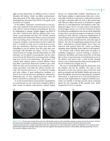
long‐term risk for stent dysfunction. The presence of a the dilated renal pelvis with a small needle, through
ureteral stent induces passive ureteral dilation which which a wire is advanced under fluoroscopic guidance to
may allow a previously obstructive urolith to pass, after the site of obstruction (Figure 122.12).
which the stent can be removed via an outpatient cysto The wire is manipulated through the obstruction, into
scopic procedure. Currently, the authors monitor ani the bladder, and out the urethra. The stent is then
mals with ureteral stents every 3–4 months by imaging advanced over this wire and into place within the renal
and urine culture. If stent malfunction develops, evi pelvis proximally and the bladder distally and urine now
denced by recurrent infection, pollakiuria, obstruction, flows through the stent lumen, bypassing the neoplastic
abdominal pain, or stent migration/fracture, then the obstruction. A small series of 12 cases found that percu
stent is removed via cystoscopy or cystotomy. taneous stent implantation for trigonal urothelial carci
To avoid eventual dysuria and potential complications noma was feasible in all dogs and the stents remained
of a ureteral stent in cats, or for those animals in which a patent for the remainder of the animals’ lives, which
stent cannot be placed, subcutaneous ureteral bypass ranged from 0.25 to 11 months.
(a) (b) (c)
Figure 122.12 Fluoroscopic images from an 8‐year‐old Labrador retriever with urothelial neoplasia causing ureteral obstruction. (a) Direct
puncture of the renal pelvis was obtained and a wire was advanced down the dilated ureter to the site of obstruction at the
ureterovesicular junction. (b) With manipulation, the wire is advanced across the obstruction and into the bladder. (c) The stent has been
positioned throughout the length of the ureter with one pigtail in the renal pelvis and one in the bladder, allowing restoration of urine
flow past the malignant obstruction.