
Page 1207 - Clinical Small Animal Internal Medicine
P. 1207
123 Urolithiasis in Small Animals 1145
(a) (b)
VetBooks.ir
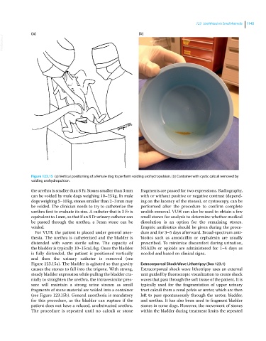
Figure 123.15 (a) Vertical positioning of a female dog to perform voiding urohydropulsion. (b) Container with cystic calculi removed by
voiding urohydropulsion.
the urethra is smaller than 8 Fr. Stones smaller than 3 mm fragments are passed for two expressions. Radiography,
can be voided by male dogs weighing 10–25 kg. In male with or without positive or negative contrast (depend
dogs weighing 5–10 kg, stones smaller than 2–3 mm may ing on the lucency of the stones), or cystoscopy, can be
be voided. The clinician needs to try to catheterize the performed after the procedure to confirm complete
urethra first to evaluate its size. A catheter that is 3 Fr is urolith removal. VUH can also be used to obtain a few
equivalent to 1 mm, so that if an 8 Fr urinary catheter can small stones for analysis to determine whether medical
be passed through the urethra, a 3 mm stone can be dissolution is an option for the remaining stones.
voided. Empiric antibiotics should be given during the proce
For VUH, the patient is placed under general anes dure and for 3–5 days afterward. Broad‐spectrum anti
thesia. The urethra is catheterized and the bladder is biotics such as amoxicillin or cephalexin are usually
distended with warm sterile saline. The capacity of prescribed. To minimize discomfort during urination,
the bladder is typically 10–15 mL/kg. Once the bladder NSAIDs or opioids are administered for 1–4 days as
is fully distended, the patient is positioned vertically needed and based on clinical signs.
and then the urinary catheter is removed (see
Figure 123.15a). The bladder is agitated so that gravity Extracorporeal Shock Wave Lithotripsy (Box 123.1)
causes the stones to fall into the trigone. With strong, Extracorporeal shock wave lithotripsy uses an external
steady bladder expression while pulling the bladder cra unit guided by fluoroscopic visualization to create shock
nially to straighten the urethra, the intravesicular pres waves that pass through the soft tissue of the patient. It is
sure will maintain a strong urine stream as small typically used for the fragmentation of upper urinary
fragments of stone material are voided into a container tract calculi from a renal pelvis or ureter, which are then
(see Figure 123.15b). General anesthesia is mandatory left to pass spontaneously through the ureter, bladder,
for this procedure, as the bladder can rupture if the and urethra. It has also been used to fragment bladder
patient does not have a relaxed, unobstructed urethra. stones in some dogs. However, the movement of stones
The procedure is repeated until no calculi or stone within the bladder during treatment limits the repeated