
Page 1208 - Clinical Small Animal Internal Medicine
P. 1208
1146 Section 10 Renal and Genitourinary Disease
shock wave effect and may result in larger than desired
Box 123.1 Candidates for extracorporeal shock wave
VetBooks.ir lithotripsy fragments. After ESWL for bladder stones, VUH or cys
toscopic stone basket retrieval is required for stone frag
ment removal. Typically, intracorporeal methods are
Nephroliths (dogs)
Indications preferred for optimal fragmentation and immediate
removal of cystoliths.
Hydronephrosis
●
Recurrent infection Intracorporeal Lithotripsy
●
Pain Electrohydraulic lithotripsy (EHL) and laser (Ho:YAG:
●
Worsening chronic renal failure holmium:yttrium, aluminum, garnet) lithotripsy were
●
Stone size developed in the 1970s to fragment bladder uroliths in
<10 mm: ESWL alone humans, with success rates reported to exceed 90%. Both
●
>10–25 mm: ESWL with ureteral stent or PCNL types of intracorporeal lithotripsy have been described
●
>25 mm: consider PCNL in veterinary patients. To date, the Ho:YAG laser is the
●
most commonly used device for intracorporeal litho
Ureteroliths (dogs and cats) tripsy in veterinary and human medicine.
During laser lithotripsy, the patient is placed under
Indications general anesthesia as for routine cystoscopy. Once the
Ureteral obstruction
● urolith is visualized with the cystoscope, a small‐
Pain
● diameter flexible quartz laser fiber (200, 365, or 550 μm)
Recurrent infection
● is guided through the working channel. The tip of the
Patient characteristics fiber is placed in direct contact with the surface of the
Dogs with ureteroliths; cats with distal ureteroliths
● urolith at a 90° angle, and pulsed laser energy is trans
Normal coagulation status
● mitted from the energized crystal to the urolith via the
Not pregnant
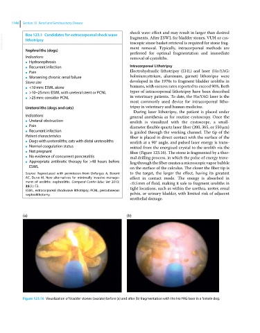
● fiber (Figure 123.16). The stone is fragmented by a ther
No evidence of concurrent pancreatitis
● mal drilling process, in which the pulse of energy trave
Appropriate antibiotic therapy for >48 hours before
● ling through the fiber creates a microscopic vapor bubble
ESWL on the surface of the calculus. The closer the fiber tip is
Source: Reproduced with permission from Defarges A, Berent to the target, the larger the effect, having its greatest
AC, Dunn M. New alternatives for minimally invasive manage- effect in contact mode. The energy is absorbed in
ment of uroliths: nephroliths. Compend Contin Educ Vet 2013; <0.5 mm of fluid, making it safe to fragment uroliths in
35(2): E3. tight locations, such as within the urethra, ureter, renal
ESWL, extracorporeal shockwave lithotripsy; PCNL, percutaneous
nephrolithotomy. pelvis, or urinary bladder, with limited risk of adjacent
urothelial damage.
(a) (b)
Figure 123.16 Visualization of bladder stones (oxalate) before (a) and after (b) fragmentation with the Ho:YAG laser in a female dog.