
Page 1526 - Clinical Small Animal Internal Medicine
P. 1526
1464 Section 12 Skin and Ear Diseases
VetBooks.ir
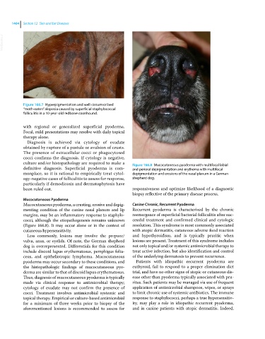
Figure 166.7 Hyperpigmentation and well‐circumscribed
“moth‐eaten” alopecia caused by superficial staphylococcal
folliculitis in a 10‐year‐old redbone coonhound.
with regional or generalized superficial pyoderma.
Focal, mild presentations may resolve with daily topical
therapy alone.
Diagnosis is achieved via cytology of exudate
obtained by rupture of a pustule or avulsion of crusts.
The presence of extracellular cocci or phagocytosed
cocci confirms the diagnosis. If cytology is negative,
culture and/or histopathology are required to make a Figure 166.8 Mucocutaneous pyoderma with multifocal labial
definitive diagnosis. Superficial pyoderma is com- and perioral depigmentation and erythema with multifocal
monplace, so it is rational to empirically treat cytol- depigmentation and erosions of the nasal planum in a German
ogy‐negative cases of folliculitis to assess for response, shepherd dog.
particularly if demodicosis and dermatophytosis have
been ruled out. responsiveness and optimize likelihood of a diagnostic
biopsy reflective of the primary disease process.
Mucocutaneous Pyoderma
Mucocutaneous pyoderma, a crusting, erosive and depig- Canine Chronic, Recurrent Pyoderma
menting condition of the canine nasal planum and lip Recurrent pyoderma is characterized by the chronic
margins, may be an inflammatory response to staphylo- reemergence of superficial bacterial folliculitis after suc-
cocci, although the etiopathogenesis remains unknown cessful treatment and confirmed clinical and cytologic
(Figure 166.8). It may occur alone or in the context of resolution. This syndrome is most commonly associated
cutaneous hypersensitivity. with atopic dermatitis, cutaneous adverse food reaction
Less commonly, lesions may involve the prepuce/ and hypothyroidism, and is typically pruritic when
vulva, anus, or eyelids. Of note, the German shepherd lesions are present. Treatment of this syndrome includes
dog is overrepresented. Differentials for this condition not only topical and/or systemic antimicrobial therapy to
include discoid lupus erythematosus, pemphigus folia- treat active infection, but also identification and control
ceus, and epitheliotropic lymphoma. Mucocutaneous of the underlying dermatosis to prevent recurrence.
pyoderma may occur secondary to these conditions, and Patients with idiopathic recurrent pyoderma are
the histopathologic findings of mucocutaneous pyo- euthyroid, fail to respond to a proper elimination diet
derma are similar to that of discoid lupus erythematosus. trial, and have no other signs of atopic or cutaneous dis-
Thus, diagnosis of mucocutaneous pyoderma is typically ease other than pyoderma typically associated with pru-
made via clinical response to antimicrobial therapy; ritus. Such patients may be managed via use of frequent
cytology of exudate may not confirm the presence of application of antimicrobial shampoos, wipes, or sprays
cocci. Treatment involves antimicrobial systemic and to limit chronic use of systemic antibiotics. The immune
topical therapy. Empirical or culture‐based antimicrobial response to staphylococci, perhaps a true hypersensitiv-
for a minimum of three weeks prior to biopsy of the ity, may play a role in idiopathic recurrent pyoderma,
aforementioned lesions is recommended to assess for and in canine patients with atopic dermatitis. Indeed,