
Page 1529 - Clinical Small Animal Internal Medicine
P. 1529
166 Bacterial Pyodermas 1467
may be similarly used as a foot soak for drying and antimi- The most common primary factors are atopic der-
VetBooks.ir crobial effects. Antimicrobial wipes and sprays may be matitis and cutaneous adverse food reaction, as well as
concurrent behavioral etiologies. Importantly, the
considered, but acid‐ and alcohol‐based products should
be avoided in erosive and ulcerative presentations (see
staphylococcal pyoderma. This must be treated to
“Treatment of pyodermas” section for additional treat- most common perpetuating factor is secondary deep
ment recommendations). reduce lesion size and severity. Common bacterial gen-
Focal nodular staphylococcal or mixed bacterial lesions era concurrently isolated from ALD lesions include
may be treated with silver sulfadiazine cream, clindamy- Pseudomonas and Enterobacter. Importantly, methicil-
cin ointment, or mupirocin ( reserved for resistant staph- lin‐resistant and multidrug‐resistant staphylococcal
ylococcal infections). Application of dimethyl sulfoxide infections are common in ALD. Cultures obtained
(DMSO) just before or after application of the topical from the surface of the lesions do not accurately repre-
antimicrobial may improve its penetration and provide sent the organisms within the lesion. Thus, tissue
additional antiinflammatory effects. Topical corticoster- biopsy for culture and sensitivity is indicated to select
oid treatment (as the sole ingredient or a combination antibiotic therapy, as treatment required is typically in
product) may be helpful for additional antiinflammatory excess of eight weeks.
benefits, particularly for hypersensitivity. Topical corti-
costeroids should be avoided in the presence of come-
dones and milia, as they may exacerbate these lesions.
Systemic antimicrobial therapy is indicated for nodules, Methicillin‐Resistant and Multidrug‐
draining tracts or severe bacterial surface pyoderma (see Resistant Staphylococci
“Treatment of pyodermas” section). Deep pyoderma
(draining nodules, crusts, ulcerations) typically require a Antimicrobial resistance in veterinary medicine has
minimum of six weeks of systemic antimicrobial therapy, rapidly evolved in recent years from a mere theoretical
and should be continued four weeks beyond cytologic possibility to an unwelcome reality. Of concern, methi-
and clinical resolution (including palpation). cillin‐resistant (MR) and multidrug‐resistant (MDR)
staphylococci pose a risk of difficult‐to‐treat nosoco-
Acral Lick Dermatitis mial infections and, to a lesser extent, zoonotic infec-
Although commonly encountered in canine practice, acral tion. To best serve our community and patients in
lick dermatitis (ALD) is a challenging condition to manage the present era of antimicrobial resistance, critical
given its multifactorial genesis and chronic nature. examination of infection control and antimicrobial
Optimal control is achieved via identification and control prescribing practices as individuals and as a profession
of contributing factors. Acral lick dermatitis is character- is essential.
ized by repetitive licking, typically of the distal limbs. Methicillin‐resistant staphylococci possess the mecA
Lesions, commonly known as lick granulomas, take the genetic element, encoding penicillin binding protein
form of erosive to ulcerative nodules or plaques with 2a (PBP2a). Beta‐lactam antibiotics cannot bind to
purulent and/or hemorrhagic exudates (Figure 166.13). PBP2a, rendering them ineffective. The mecA gene is
present within staphylococcal chromosomal cassettes
(SCCs) that are also horizontally transferred amongst
Staphylococcus spp. Staphylococcal chromosomal cas-
settes may contain other antimicrobial resistance genes,
imparting multidrug resistance in nearly all strains of
MR staphylococci encountered in veterinary practice. Of
note, beta‐lactam antimicrobial use selects for survival
of MR staphylococci, but does not create, de novo, spon-
taneous resistance.
Cutaneous MR staphylococcal infection and coloni-
zation are a worldwide phenomenon, recognized in the
dog, cat, horse, cattle, and swine. In years past, MR
staphylococcus appeared to be very rare to absent in
the canine and feline population but a remarkable
increase in frequency of methicillin resistance in the
canine and feline population was noted early in the
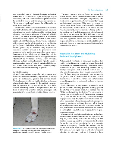
Figure 166.13 Acral lick granuloma with erosive surface on the
carpus of a 9‐year‐old Labrador mix; deep staphylococcal 21st century. The rapid rise of MR staphylococcal
pyoderma and atopic dermatitis were contributing factors. infection in veterinary patients is thought to have