
Page 1534 - Clinical Small Animal Internal Medicine
P. 1534
1472 Section 12 Skin and Ear Diseases
VetBooks.ir Box 167.1 Primary causes for otitis externa Box 167.2 Predisposing factors in otitis externa
Allergy – atopic dermatitis, cutaneous adverse food
Conformation of the ear – narrowed canal (shar‐pei),
reaction, contact allergy
concave pinna (spaniels), large pendulous pinnae
Endocrine disease – hypothyroidism (dog), hyperthy- hairy external ear canal (spaniels, poodles), hairy
roidism (cat), hyperadrenocorticism (dog and cat) ( basset hounds, bloodhounds)
Ectoparasites – Otodectes cynotis, Demodex spp., Inappropriate cleaning, irritant treatment – use of
Otobius megnini astringent, low‐acidity topical products or potent
Autoimmune/immune-mediated disease – pemphigus ceruminolytic cleaner when not appropriate
foliaceus, discoid lupus erythematosus, erythema Wetting of the ear – swimming dogs, water‐based
multiforme, juvenile cellulitis, drug reactions, vasulitis cleaners, topical medication mixed in water
Keratinization disorders – sebaceous adenitis, zinc‐ Obstructive ear disease – polyps, ceruminous gland
responsive dermatosis, primary idiopathic seborrhea adenomas, other neoplasia
Foreign bodies – grass awns, ceruminoliths
( accumulations of cerumen due to failure of epithelial
migration)
Box 167.3 Perpetuating factors in otitis externa
Chronic changes – epidermal and glandular hyperpla-
sia, stenosis, failure of epidermal migration, calcifica-
tion of ear canals, ulceration
Otitis media
pendulous pinnae such as spaniels, basset hounds, and
bloodhounds are predisposed to disease, as are breeds
such as the shar‐pei which have narrowed canals.
History and Clinical Signs
The key aims of history taking are to try to establish the
primary cause for the otitis and any predisposing, per-
petuating or secondary factors that may be contributing
to the disease. Primary causes (see Box 167.1) should be
identified in all cases of otitis. Successful management of
these triggers is particularly important in chronic dis-
ease. Predisposing factors, which are listed in Box 167.2,
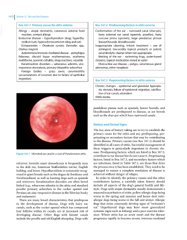
Figure 167.1 Ulcerated ear canal in a case of Pseudomonas otitis.
contribute to ear disease but do not cause it. Perpetuating
factors, listed in Box 167.3, and secondary factors which
retriever. Juvenile‐onset demodicosis is frequently seen are infections, listed in Table 167.1, are those that drive
in the shih tzu, American Staffordshire terrier, English the process once it has been established. They need to be
bulldog, and boxer. Hypothyroidism is commonly recog- managed to ensure a complete resolution of disease is
nized in giant breeds such as the dogue de Bordeaux and achieved without danger of relapse.
Newfoundland, as well as hunting dogs such as spaniels In order to identify the primary causes and the other
and retrievers. Keratinization disorders are often breed contributory factors, a carefully taken history should
linked (e.g., sebaceous adenitis in the akita and standard include all aspects of the dog’s general health and life-
poodle; primary seborrhea in the cocker spaniel and style. Dogs with atopic dermatitis usually demonstrate a
Persian cat; zinc‐responsive disease in the Siberian husky seasonal exacerbation of otitis, pollen‐allergic dogs being
and malamute). worse in the spring and summer and house dust mite‐
There are many breed characteristics that predispose allergic dogs being worse in the fall and winter. Allergic
to the development of disease. Dogs with hairy ear dogs that swim commonly develop signs of “swimmer’s
canals, such as the cocker spaniel which has compound ear.” Hypothyroid dogs may have more generalized
hair follicles within its canals, are at increased risk of systemic signs such as lethargy and a poor exercise toler-
developing disease. Other dogs with hirsute canals ance. Where otitis has an acute onset and the disease
include the poodle and old English sheepdog. Dogs with progresses rapidly to become severe, immune‐mediated