
Page 1536 - Clinical Small Animal Internal Medicine
P. 1536
1474 Section 12 Skin and Ear Diseases
VetBooks.ir
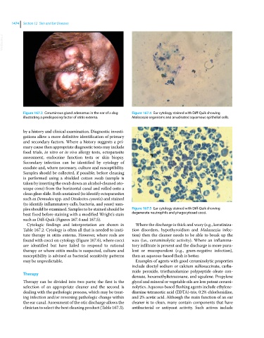
Figure 167.3 Ceruminous gland adenomas in the ear of a dog Figure 167.4 Ear cytology stained with Diff‐Quik showing
illustrating a predisposing factor of otitis externa. Malassezia organisms and anucleated squamous epithelial cells.
by a history and clinical examination. Diagnostic investi-
gations allow a more definitive identification of primary
and secondary factors. Where a history suggests a pri-
mary cause then appropriate diagnostic tests may include
food trials, in vitro or in vivo allergy tests, ectoparasite
assessment, endocrine function tests or skin biopsy.
Secondary infection can be identified by cytology of
exudate and, where necessary, culture and susceptibility.
Samples should be collected, if possible, before cleaning
is performed using a shielded cotton swab (sample is
taken by inserting the swab down an alcohol‐cleaned oto-
scope cone) from the horizontal canal and rolled onto a
clean glass slide. Both unstained (to identify ectoparasites
such as Demodex spp. and Otodectes cynotis) and stained
(to identify inflammatory cells, bacteria, and yeast) sam-
ples should be examined. Samples to be stained should be Figure 167.5 Ear cytology stained with Diff‐Quik showing
heat fixed before staining with a modified Wright’s stain degenerate neutrophils and phagocytosed cocci.
such as Diff‐Quik (Figures 167.4 and 167.5).
Cytologic findings and interpretation are shown in Where the discharge is thick and waxy (e.g., keratiniza-
Table 167.2. Cytology is often all that is needed to insti- tion disorders, hypothyroidism and Malassezia infec-
tute therapy in otitis externa. However, where rods are tion) then the cleaner needs to be able to break up the
found with cocci on cytology (Figure 167.6), where cocci wax (i.e., ceruminolytic activity). Where an inflamma-
are identified but have failed to respond to rational tory infiltrate is present and the discharge is more puru-
therapy or where otitis media is suspected, culture and lent or mucopurulent (e.g., gram‐negative infection),
susceptibility is advised as bacterial sensitivity patterns then an aqueous‐based flush is better.
may be unpredictable. Examples of agents with good ceruminolytic properties
include dioctyl sodium or calcium sulfosuccinate, carba-
mide peroxide, triethanolamine polypeptide oleate con-
Therapy
densate, hexamethyltetracosane, and squalene. Propylene
Therapy can be divided into two parts; the first is the glycol and mineral or vegetable oils are less potent cerumi-
selection of an appropriate cleaner and the second is nolytics. Aqueous‐based flushing agents include ethylene-
dealing with the pathologic process, which may be treat- diamine tetraacetic acid (EDTA)‐tris, 0.2% chlorhexidine,
ing infection and/or reversing pathologic change within and 2% acetic acid. Although the main function of an ear
the ear canal. Assessment of the otic discharge allows the cleaner is to clean, many contain components that have
clinician to select the best cleaning product (Table 167.3). antibacterial or antiyeast activity. Such actives include