
Page 1535 - Clinical Small Animal Internal Medicine
P. 1535
167 Otitis 1473
Table 167.1 Secondary causes in otitis externa
VetBooks.ir Infectious organism Acute disease Chronic disease
Bacteria (most Gram‐positive bacteria Staphylococcus spp. Enterococcus spp.
common isolates) Streptococcus spp. Corynebacteria spp.
Gram‐negative bacteria Uncommon Pseudomonas spp. Proteus spp.
Escherichia coli
Anaerobic bacteria Uncommon Bacteroides spp.
Yeast Malassezia Malassezia pachydermatis
(most common isolate) pachydermatis
disease such as drug eruptions and autoimmune
disease should be considered. Contact irritant or hyper-
sensitivity should be considered when signs of otitis
become worse after commencement of topical therapy.
Contact reactions may be to one of the active ingredients
in the topical product such as an antibiotic, antifungal
or glucocorticoid, or can be to the vehicle (e.g., propyl-
ene glycol).
Examination of each case should include a full physical
and dermatologic inspection before examining the pin-
nae and ear canal. A physical examination may reveal
signs, for example of bradycardia in a hypothyroid dog.
Accompanying signs of more generalized disease may
provide useful clues. Dogs with atopic dermatitis or
adverse food reaction may have pedal salivary staining
due to foot licking, or recurrent ventral pyoderma.
Dogs with endocrine disease may have seborrhea, a poor
hair coat or bilaterally symmetric alopecia which
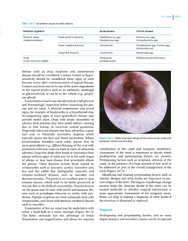
typically spares the face and distal extremities. Where Figure 167.2 Video otoscopic image of the external ear canal and
keratinization disorders cause otitis, lesions may be tympanic membrane of a dog.
more generalized (e.g., diffuse thinning of the coat with
prominent follicular casts on hairs in cases of sebaceous visualization of the canal and tympanic membrane.
adenitis). Dogs that shake their heads or traumatize their Assessment of the canal is important to decide which
pinnae without signs of otitis may be in the early stages predisposing and perpetuating factors are present.
of allergy or may have disease that principally affects Predisposing factors such as neoplasia, stenosis of the
the pinnae. These diseases include those caused by canal, or the presence of a large amount of hair need to
ectoparasites such as Sarcoptes scabiei, Notoedres cati, be addressed as part of the overall management of the
lice and the rabbit flea (Spilopsyllus cuniculi), and otitis (Figure 167.3).
immune‐mediated diseases such as vasculitis and Identifying and treating perpetuating factors such as
dermatomyositis. The pinna is an excellent area to view chronic changes and otitis media are important to pre-
primary lesions, which tend to be better preserved at vent relapse of the otitis. The degree of pathologic change
this site due to the difficult accessibility. Pustular lesions present helps the clinician decide if the otitis can be
on the pinna may be seen with sterile autoimmune dis- treated medically or whether surgical intervention is
ease such as pemphigus foliaceus or, rarely, with pyo- more appropriate. Assessment of the tympanic mem-
derma. Papular eruptions are most commonly seen with brane can help in making a diagnosis of otitis media if
ectoparasites, and ulcers with immune‐mediated disease the structure is abnormal or ruptured.
such as vasculitis.
Examination of the ear canal may be undertaken with
either a hand‐held or a video otoscope (Figure 167.2). Diagnosis
The latter obviously has the advantage of better Predisposing and perpetuating factors, and to some
illumination and magnification and allows for superior degree primary and secondary causes, can be recognized