
Page 1527 - Clinical Small Animal Internal Medicine
P. 1527
166 Bacterial Pyodermas 1465
subcutaneously injected commercial S. aureus phage atopic dermatitis or cutaneous adverse food reaction,
VetBooks.ir lysate antigens (Staphage Lysate®, Delmont Laboratories) cystic hemorrhagic, bullous lesions are often observed
on the chin and interdigital areas. Staphylococci repre-
may be used as maintenance therapy to reduce or elimi-
nate recurrence of pyoderma. This treatment is indicated
may also be accompanied by infection with Enterococcus,
in patients with idiopathic or atopic recurrent pyoderma sent the predominant organism but deep pyoderma
cases failing to respond to topical maintenance therapy. Pseudomonas, and Proteus species as well as Escherichia
coli. Differentials include dimorphic and opportunistic
mycoses, pythiosis, lagenidiosis, neoplasia, foreign
Deep Pyodermas
body, sterile pyogranulomatous dermatitis, and other
Generalized Deep Pyoderma bacterial etiologies, including species in the genera
Generalized deep pyoderma arises due to furunculosis Mycobacterium, Actinomyces, and Nocardia. Cytologic
(follicular rupture) and foreign body response directed presence of numerous extracellular cocci or phagocy-
at the liberated hair shafts in the dermis. Lesions include tosed cocci supports the diagnosis. Given the diverse
nodules, hemorrhagic bullae, ulcers, draining tracts and clinical differentials and requirement for prolonged
crusts with variable pain and pruritus (Figures 166.9 treatment, bacterial culture (aerobic and anaerobic)
and 166.10). and sensitivity, fungal culture, and histopathology are
In cats, deep pyoderma most commonly presents in recommended for definitive diagnosis and appropriate
the context of chin acne. In short‐coated breeds with treatment.
Canine Acne
This disease is typified by deep folliculitis and furuncu-
losis affecting the chin and perioral margin, and is a
focal form of deep pyoderma. Chin acne usually mani-
fests in young dogs between 3 and 12 months of age
although it often persists throughout adulthood and
may be complicated by concurrent atopic dermatitis or
cutaneous adverse food reaction. Breeds with a short,
coarse hair coat are predisposed, including boxers,
English and French bulldogs, and Boston terriers.
Lesions consist of papules and nodules that often
become erosive and ulcerative with resultant draining
tract formation. Hemorrhagic bullae are also common
(Figure 166.11).
Demodicosis should be ruled out by performing deep
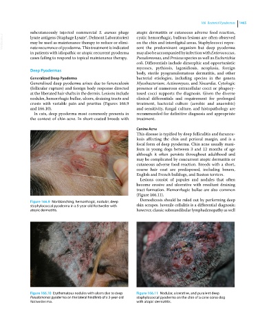
Figure 166.9 Nonblanching, hemorrhagic, nodular, deep
staphylococcal pyoderma in a 5‐year‐old Rottweiler with skin scrapes. Juvenile cellulitis is a differential diagnosis;
atopic dermatitis. however, classic submandibular lymphadenopathy as well
Figure 166.10 Erythematous nodules with ulcers due to deep Figure 166.11 Nodular, ulcerative, and purulent deep
Pseudomonas pyoderma on the lateral hindlimb of a 3‐year‐old staphylococcal pyoderma on the chin of a cane corso dog
Rottweiler mix. with atopic dermatitis.