
Page 1561 - Clinical Small Animal Internal Medicine
P. 1561
170 Miscellaneous Skin Diseases 1499
hematochezia, it was found that pancreatitis, inflamma-
VetBooks.ir tory bowel disease, and adverse food reaction were the
most common triggers, although the cause of the gastro-
intestinal signs could not be documented for all dogs. In
some dogs, an adverse drug reaction was considered as a
possible trigger.
Signalment
Labrador retrievers and their crosses appear to be over-
represented, but the disease can occur in any breed. Age
and sex predilections have not been identified.
History and Clinical Signs
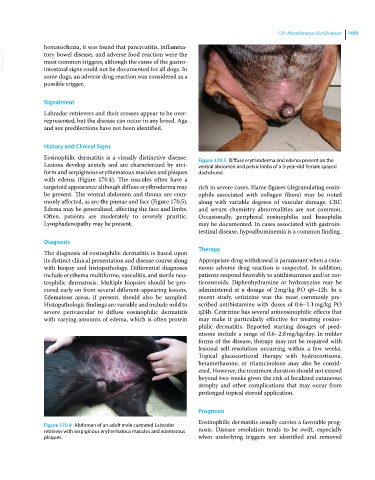
Eosinophilic dermatitis is a visually distinctive disease. Figure 170.5 Diffuse erythroderma and edema present on the
Lesions develop acutely and are characterized by arci- ventral abdomen and pelvic limbs of a 3‐year‐old female spayed
form and serpiginous erythematous macules and plaques dachshund.
with edema (Figure 170.4). The macules often have a
targetoid appearance although diffuse erythroderma may rich in severe cases. Flame figures (degranulating eosin-
be present. The ventral abdomen and thorax are com- ophils associated with collagen fibers) may be noted
monly affected, as are the pinnae and face (Figure 170.5). along with variable degrees of vascular damage. CBC
Edema may be generalized, affecting the face and limbs. and serum chemistry abnormalities are not common.
Often, patients are moderately to severely pruritic. Occasionally, peripheral eosinophilia and basophilia
Lymphadenopathy may be present. may be documented. In cases associated with gastroin-
testinal disease, hypoalbuminemia is a common finding.
Diagnosis
Therapy
The diagnosis of eosinophilic dermatitis is based upon
its distinct clinical presentation and disease course along Appropriate drug withdrawal is paramount when a cuta-
with biopsy and histopathology. Differential diagnoses neous adverse drug reaction is suspected. In addition,
include erythema multiforme, vasculitis, and sterile neu- patients respond favorably to antihistamines and/or cor-
trophilic dermatosis. Multiple biopsies should be pro- ticosteroids. Diphenhydramine or hydroxyzine may be
cured early on from several different‐appearing lesions. administered at a dosage of 2 mg/kg PO q8–12h. In a
Edematous areas, if present, should also be sampled. recent study, cetirizine was the most commonly pre-
Histopathologic findings are variable and include mild to scribed antihistamine with doses of 0.6–1.1 mg/kg PO
severe perivascular to diffuse eosinophilic dermatitis q24h. Cetirizine has several antieosinophilic effects that
with varying amounts of edema, which is often protein may make it particularly effective for treating eosino-
philic dermatitis. Reported starting dosages of pred-
nisone include a range of 0.6–2.8 mg/kg/day. In milder
forms of the disease, therapy may not be required with
lesional self‐resolution occurring within a few weeks.
Topical glucocorticoid therapy with hydrocortisone,
betamethasone, or triamcinolone may also be consid-
ered. However, the treatment duration should not extend
beyond two weeks given the risk of localized cutaneous
atrophy and other complications that may occur from
prolonged topical steroid application.
Prognosis
Eosinophilic dermatitis usually carries a favorable prog-
Figure 170.4 Abdomen of an adult male castrated Labrador
retriever with serpiginous erythematous macules and edematous nosis. Disease resolution tends to be swift, especially
plaques. when underlying triggers are identified and removed