
Page 1563 - Clinical Small Animal Internal Medicine
P. 1563
170 Miscellaneous Skin Diseases 1501
other breeds reported to date have had the nonepider- Golden Retriever
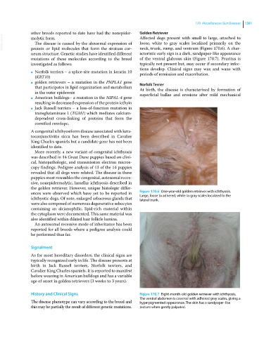
VetBooks.ir molytic form. loose, white to gray scales localized primarily on the
Affected dogs present with small to large, attached to
The disease is caused by the abnormal expression of
protein or lipid molecules that form the stratum cor-
neck, trunk, rump, and ventrum (Figure 170.6). A char-
neum structure. Genetic studies have identified different acteristic early sign is a dark, sandpaper‐like appearance
mutations of these molecules according to the breed of the ventral glabrous skin (Figure 170.7). Pruritus is
investigated as follows: typically not present but, may occur if secondary infec-
tions develop. Clinical signs may wax and wane with
Norfolk terriers – a splice‐site mutation in keratin 10
● periods of remission and exacerbation.
(KRT10)
golden retrievers – a mutation in the PNPLA1 gene
● Norfolk Terrier
that participates in lipid organization and metabolism At birth, the disease is characterized by formation of
in the outer epidermis superficial bullae and erosions after mild mechanical
American bulldogs– a mutation in the NIPAL‐4 gene
●
resulting in decreased expression of the protein icthyin
Jack Russell terriers – a loss‐of‐function mutation in
●
transglutaminase 1 (TGM1) which mediates calcium‐
dependent cross‐linking of proteins that form the
cornified envelope.
A congenital ichthyosiform disease associated with kera-
toconjunctivitis sicca has been described in Cavalier
King Charles spaniels but a candidate gene has not been
identified to date.
More recently, a new variant of congenital ichthyosis
was described in 14 Great Dane puppies based on clini-
cal, histopathologic, and transmission electron micros-
copy findings. Pedigree analysis of 13 of the 14 puppies
revealed that all dogs were related. The disease in these
puppies most resembles the congenital, autosomal reces-
sive, nonepidermolytic, lamellar ichthyosis described in
the golden retriever. However, unique histologic differ-
ences were observed which have yet to be reported in Figure 170.6 One‐year‐old golden retriever with ichthyosis.
Large, loose to adhered, white to gray scales localized to the
ichthyotic dogs. Of note, enlarged sebaceous glands that lateral trunk.
were also composed of numerous degenerative sebocytes
containing an alcianophilic, lipid‐rich material within
the cytoplasm were documented. This same material was
also identified within dilated hair follicle lumina.
An autosomal recessive mode of inheritance has been
reported for all breeds where a pedigree analysis could
be performed thus far.
Signalment
As for most hereditary disorders, the clinical signs are
typically recognized early in life. The disease presents at
birth in Jack Russell terriers, Norfolk terriers, and
Cavalier King Charles spaniels. It is reported to manifest
before weaning in American bulldogs and has a variable
age of onset in golden retrievers (3 weeks to 3 years).
History and Clinical Signs Figure 170.7 Eight‐month‐old golden retriever with ichthyosis.
The ventral abdomen is covered with adhered gray scales, giving a
The disease phenotype can vary according to the breed and hyperpigmented appearance. The skin has a sandpaper‐like
this may be partially the result of different genetic mutations. texture when gently palpated.