
Page 1565 - Clinical Small Animal Internal Medicine
P. 1565
170 Miscellaneous Skin Diseases 1503
ichthyosis was documented in a goldendoodle dog based food reaction, atopic dermatitis, flea bite allergy or mos-
VetBooks.ir on histopathologic findings and genetic testing, which quito bite hypersensitivity, suggesting an immunologic
dysregulation. The role of bacteria in lesion development
identified the PNPLA1 mutation found in golden retriev-
ers, indicating that the disease was likely inherited from
significantly or resolved with antibiotic therapy. Many
its golden retriever predecessors. is still questionable but certain lesions have improved
cases remain idiopathic and some cats may be genetically
predisposed to develop this condition. Cats can have a
Diagnosis single lesion, multiples of the same type or a combina-
The diagnosis historically has been based on the charac- tion of different ones. Lesions may be refractory to treat-
teristic history, clinical signs, and histopathologic find- ment, resolve with or without treatment and not return,
ings. When performed, skin biopsies should be submitted or be recurrent.
to a pathologist with experience in dermatology. A com-
mercially available genetic test is now offered through Signalment
Optigen (www.optigen.com/) and is easily performed
using a venous blood sample and has become the authors’ There is no breed, sex or age predilection but linear
primary means of confirming a diagnosis of ichthyosis in granuloma lesions typically develop in cats less than
breeds where testing is an option. Electron microscopy 2 years of age.
and genetic studies are needed to characterize the
molecular abnormality and mode of transmission. History and Clinical Signs
Lesions associated with the complex include eosinophilic
Therapy granuloma, eosinophilic plaque, and indolent ulcer.
The treatment goals should be to prevent/treat secondary
infections and reduce the excessive scaling, hyperkerato- Eosinophilic Granuloma
sis, and inflammation by restoring the stratum corneum This is the only true granulomatous lesion of the complex
barrier. This may be achieved by various topical therapies and has various clinical presentations such as nodules,
which should be tailored to the patient’s needs. Shampoos papules or linear plaques. They can be single or multiple
with keratolytic and keratoplastic properties such as sul- and develop anywhere on the body, including the lips
fur and salicylic acid, followed by a good moisturizer, can (pouting lip), chin (fat chin), oral cavity, footpads, pinnae,
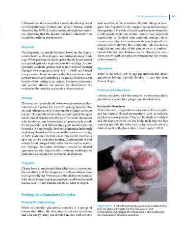
be used 2–3 times weekly. Products containing lipids such caudal aspect of thighs or other areas (Figure 170.11).
as phytosphingosine (Douxo seborrhea spot‐on or spray),
or fatty acids and essential oils (Dermoscent Essential 6
spot‐on) can be used after bathing. Combinations of oral
omega‐3 and omega‐6 fatty acids can be used as adjunc-
tive therapy. Secondary infections should be treated
appropriately with topical and/or systemic antifungals or
antibiotics as required for each individual patient.
Prognosis
Clients have to understand that ichthyosis is a noncura-
ble condition and the prognosis to achieve disease con-
trol is generally fair. Veterinarians should become familiar
with the different clinical presentations and breed‐related
disease severity and educate clients on what to expect.
Eosinophilic Granuloma Complex
Etiology/Pathophysiology
Feline eosinophilic granuloma complex is a group of Figure 170.11 A cat with eosinophilic granuloma localized to the
feet. The digits of the right hind foot are swollen and
lesions that affect the skin, mucocutaneous junctions, erythematous. The footpad of the third digit is also swollen and
and oral cavity. They can develop in cats with adverse has a focal area of erosion to ulceration.