
Page 662 - Clinical Small Animal Internal Medicine
P. 662
630 Section 6 Gastrointestinal Disease
Further blood tests are indicated if none of the following
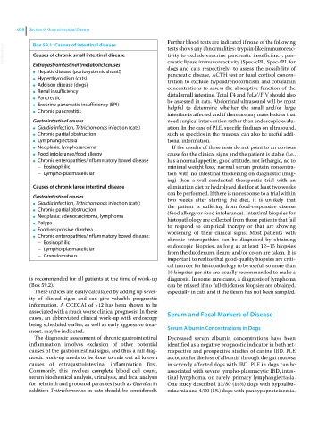
Box 59.1 Causes of intestinal disease
VetBooks.ir Causes of chronic small intestinal disease tests shows any abnormalities: trypsin‐like immunoreac-
tivity to exclude exocrine pancreatic insufficiency, pan-
creatic lipase immunoreactivity (Spec‐cPL, Spec‐fPL for
Extragastrointestinal (metabolic) causes
Hepatic disease (portosystemic shunt!) dogs and cats respectively) to assess the possibility of
● pancreatic disease, ACTH test or basal cortisol concen-
Hyperthyroidism (cats)
● tration to exclude hypoadrenocorticism and cobalamin
Addison disease (dogs)
● concentrations to assess the absorptive function of the
Renal insufficiency
● distal small intestine. Total T4 and FeLV/FIV should also
Pancreatic
● be assessed in cats. Abdominal ultrasound will be most
Exocrine pancreatic insufficiency (EPI)
● helpful to determine whether the small and/or large
Chronic pancreatitis
●
intestine is affected and if there are any mass lesions that
Gastrointestinal causes need surgical intervention rather than endoscopic evalu-
Giardia infection, Tritrichomonas infection (cats) ation. In the case of PLE, specific findings on ultrasound,
●
Chronic partial obstruction such as speckles in the mucosa, can also be useful addi-
●
Lymphangiectasia tional information.
●
Neoplasia: lymphosarcoma If the results of these tests do not point to an obvious
●
Food intolerance/food allergy cause for the clinical signs and the patient is stable (i.e.,
●
Chronic enteropathies/inflammatory bowel disease has a normal appetite, good attitude, not lethargic, no to
●
– Eosinophilic minimal weight loss, normal serum protein concentra-
– Lympho‐plasmacellular tion with no intestinal thickening on diagnostic imag-
ing) then a well‐conducted therapeutic trial with an
Causes of chronic large intestinal disease elimination diet or hydrolyzed diet for at least two weeks
can be performed. If there is no response to a trial within
Gastrointestinal causes two weeks after starting the diet, it is unlikely that
Giardia infection, Tritrichomonas infection (cats)
● the patient is suffering from food‐responsive disease
Chronic partial obstruction
● (food allergy or food intolerance). Intestinal biopsies for
Neoplasia: adenocarcinoma, lymphoma
● histopathology are collected from those patients that fail
Polyps
● to respond to empirical therapy or that are showing
Food‐responsive diarrhea
● worsening of their clinical signs. Most patients with
Chronic enteropathies/inflammatory bowel disease:
● chronic enteropathies can be diagnosed by obtaining
– Eosinophilic endoscopic biopsies, as long as at least 12–15 biopsies
– Lympho‐plasmacellular
– Granulomatous from the duodenum, ileum, and/or colon are taken. It is
important to realize that good‐quality biopsies are criti-
cal in order for histopathology to be useful, so more than
10 biopsies per site are usually recommended to make a
is recommended for all patients at the time of work‐up diagnosis. In some rare cases, a diagnosis of lymphoma
(Box 59.2). can be missed if no full‐thickness biopsies are obtained,
These indices are easily calculated by adding up sever- especially in cats and if the ileum has not been sampled.
ity of clinical signs and can give valuable prognostic
information. A CCECAI of >12 has been shown to be
associated with a much worse clinical prognosis. In these Serum and Fecal Markers of Disease
cases, an abbreviated clinical work‐up with endoscopy
being scheduled earlier, as well as early aggressive treat-
ment, may be indicated. Serum Albumin Concentrations in Dogs
The diagnostic assessment of chronic gastrointestinal Decreased serum albumin concentrations have been
inflammation involves exclusion of other potential identified as a negative prognostic indicator in both ret-
causes of the gastrointestinal signs, and thus a full diag- rospective and prospective studies of canine IBD. PLE
nostic work‐up needs to be done to rule out all known accounts for the loss of albumin through the gut mucosa
causes of extragastrointestinal inflammation first. in severely affected dogs with IBD. PLE in dogs can be
Commonly, this involves complete blood cell count, associated with severe lympho‐plasmacytic IBD, intes-
serum biochemical analysis, urinalysis, and fecal analysis tinal lymphoma, or, rarely, primary lymphangiectasia.
for helminth and protozoal parasites (such as Giardia; in One study described 12/80 (16%) dogs with hypoalbu-
addition Tritrichomonas in cats should be considered). minemia and 4/80 (5%) dogs with panhypoproteinemia.