
Page 924 - Clinical Small Animal Internal Medicine
P. 924
862 Section 9 Infectious Disease
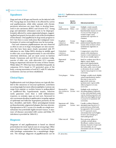
Signalment Table 83.1 Papillomavirus‐associated disease in domestic
VetBooks.ir Dogs and cats of all ages and breeds can be infected with Species
dogs and cats
PVs. Young dogs are most likely to be affected by canine
oral papillomatosis, while older animals with chronic Disease affected Clinical presentation
persistent infections are more likely to develop bowe Oral Canine Multiple, raised, smooth
noid in situ carcinoma (BISC) and invasive SCC. Young papillomatosis Feline – rare nodular to pedunculated,
pugs and miniature schnauzers seem to be dispropor hyperkeratotic masses on oral
tionately affected by canine pigmented plaques, suggest mucosa and lips (dogs), or
ing that host genetic factors play a role in the development ventral tongue (cats);
spontaneous regression is
of this disease. Oral papillomas in dogs rarely progress to common
SCC but there is some indication that malignant trans Exophytic Canine Single to multiple
formation of canine oral papillomas may be increasing. cutaneous Feline – rare pedunculated, hyperkeratotic
Oral and cutaneous papillomatoses are not observed papillomatosis masses often on head or feet;
as often in cats as in dogs; viral plaques are also uncom spontaneous regression is
mon but have been more clearly associated with PV common
infections in cats. Feline BISCs develop in middle‐aged Endophytic Canine Unpigmented, raised firm
to older cats, occur disproportionately in cats with feline cutaneous masses with central pores
retrovirus infections, and are most severe in hairless papillomatosis found on ventral abdomen or
feet; do not usually regress
breeds. Cutaneous and oral SCCs are common malig
nancies of older cats, with ultraviolet (UV) exposure Pigmented Canine Small to medium‐sized flat to
plaques
slightly raised, darkly
being an important risk factor for some of these lesions. pigmented, hyperkeratotic
While feline PV DNA has been identified frequently in masses found in clusters on
cutaneous SCCs found in UV‐protected areas of the the limbs, axilla, ventral trunk
body, a strong association of oral SCC with PV infection or abdomen; do not usually
in domestic cats has not been established. regress; may progress to in situ
carcinoma
Viral plaques Feline Multiple variably sized, slightly
Clinical Signs raised, hyperkeratotic masses
that may or may not be
pigmented; some may
Papillomatosis and viral plaque lesions are typically lim spontaneously regress while
ited to the cutaneous or mucosal epidermis, sometimes others progress to in situ
occurring singly but more often in multiples. Lesions can carcinoma
range from papular or nodular lesions to pedunculated Bowenoid in Feline Multiple scaly, crusting to
or cauliflower‐like, hyperkeratotic masses. Infection situ carcinoma Canine – rare ulcerating plaques frequently
rarely generates more than a mild inflammatory (BISC) on face, neck and limbs; some
response, so systemic signs are not seen. Bowenoid in may be slowly progressive and
others become highly invasive
situ carcinomas develop as multicentric irregular regions or progress to squamous cell
of epidermal and follicular hyperplasia primarily on the carcinoma
face, shoulders, and limbs. These premalignant lesions Squamous cell Feline Usually a solitary flattened
are hyperkeratotic, pigmented plaques that may ulcerate. carcinoma Canine – rare ulcerated mass found in UV‐
If progression to SCC occurs, it is usually as a single non (SCC) protected areas of the body
metastatic but potentially highly invasive tumor. A sum (as opposed to SCCs caused
mary of diseases associated with canine and feline PV by UV exposure); rarely
metastasize but can be highly
infections is provided in Table 83.1. invasive and damaging to
surrounding tissue
Diagnosis Feline sarcoid Feline Slow‐growing, solitary to
multiple, smooth dense
fibroblastic nodules that may
Diagnosis of oral papillomatosis is based on clinical ulcerate, located on head, neck
presentation for most typical cases in young dogs. Other and extremities; do not
types of lesions require full‐thickness excisional biopsy metastasize and are not highly
with histologic examination for a morphologic diagn invasive
osis. The etiologic diagnosis of papillomatosis can be UV, ultraviolet.