
Page 153 - Feline diagnostic imaging
P. 153
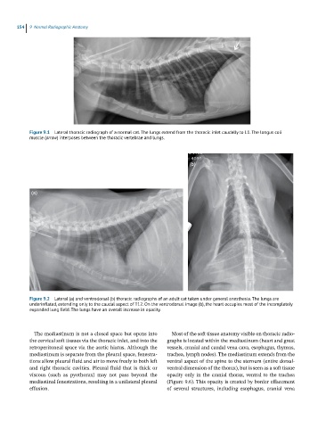
154  9  Normal Radiographic Anatomy


            Figure 9.1  Lateral thoracic radiograph of a normal cat. The lungs extend from the thoracic inlet caudally to L1. The longus coli
            muscle (arrow) interposes between the thoracic vertebrae and lungs.


                                                                       (b)


             (a)


            Figure 9.2  Lateral (a) and ventrodorsal (b) thoracic radiographs of an adult cat taken under general anesthesia. The lungs are
            underinflated, extending only to the caudal aspect of T12. On the ventrodorsal image (b), the heart occupies most of the incompletely
            expanded lung field. The lungs have an overall increase in opacity.


              The mediastinum is not a closed space but opens into   Most of the soft tissue anatomy visible on thoracic radio-
            the cervical soft tissues via the thoracic inlet, and into the   graphs is located within the mediastinum (heart and great
            retroperitoneal space via the aortic hiatus. Although the   vessels, cranial and caudal vena cava, esophagus, thymus,
            mediastinum is separate from the pleural space, fenestra-  trachea, lymph nodes). The mediastinum extends from the
            tions allow pleural fluid and air to move freely to both left   ventral aspect of the spine to the sternum (entire dorsal‐
            and right thoracic cavities. Pleural fluid that is thick or   ventral dimension of the thorax), but is seen as a soft tissue
            viscous  (such  as  pyothorax)  may  not  pass  beyond  the   opacity only in the cranial thorax, ventral to the trachea
            mediastinal fenestrations, resulting in a unilateral pleural   (Figure 9.6). This opacity is created by border effacement
            effusion.                                         of  several  structures,  including  esophagus,  cranial  vena