
Page 158 - Feline diagnostic imaging
P. 158
9.11 Normal Vascular Anatomy 159
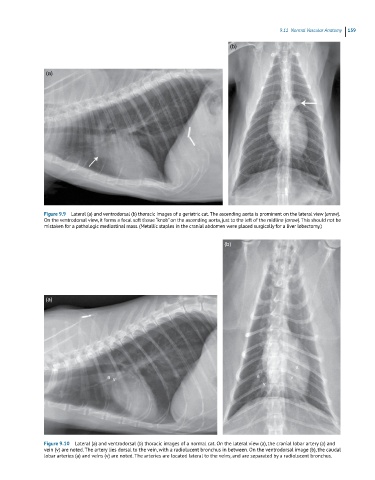
(b)
(a)
Figure 9.9 Lateral (a) and ventrodorsal (b) thoracic images of a geriatric cat. The ascending aorta is prominent on the lateral view (arrow).
On the ventrodorsal view, it forms a focal soft tissue “knob” on the ascending aorta, just to the left of the midline (arrow). This should not be
mistaken for a pathologic mediastinal mass. (Metallic staples in the cranial abdomen were placed surgically for a liver lobectomy.)
(b)
(a)
a
a v a v
v
Figure 9.10 Lateral (a) and ventrodorsal (b) thoracic images of a normal cat. On the lateral view (a), the cranial lobar artery (a) and
vein (v) are noted. The artery lies dorsal to the vein, with a radiolucent bronchus in between. On the ventrodorsal image (b), the caudal
lobar arteries (a) and veins (v) are noted. The arteries are located lateral to the veins, and are separated by a radiolucent bronchus.