
Page 156 - Feline diagnostic imaging
P. 156
9.9 eart 157
(b)
(a)
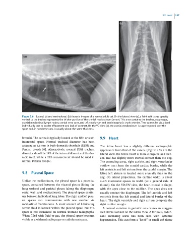
Figure 9.6 Lateral (a) and ventrodorsal (b) thoracic images of a normal adult cat. On the lateral view (a), a faint soft tissue opacity
ventral to the trachea represents the thicker portion of the cranial mediastinum (arrow). This area contains the trachea, esophagus,
cranial mediastinal lymph nodes, cranial vena cava, and left subclavian and brachiocephalic trunk arteries. They cannot be visualized
individually due to border effacement and lack of contrast. On the VD view (b), the cranial mediastinum is superimposed over the
spine and, in nonobese cats, is usually about the same thickness.
bronchi. The carina is typically located at the fifth or sixth 9.9 Heart
intercostal space. Normal tracheal diameter has been
assessed as 5.5 mm in both domestic shorthair (DSH) and The feline heart has a slightly different radiographic
Persian breeds [6]. Alternatively, normal DSH tracheal appearance from that of the canine (Figure 9.9). On the
diameter should be 18% of the internal diameter of the tho- lateral view, the feline heart is more elongated and slen-
racic inlet, while a 20% measurement should be used in der, and has slightly more sternal contact than the dog.
normal Persian cats [6]. The ascending aorta, right auricle, and right ventricular
outflow tract form the cranial cardiac border, while the
left ventricle and left atrium form the caudal margin. The
9.8 Pleural Space feline left atrium is located more cranially than in the
dog. On lateral projections, the cardiac width is about
Unlike the mediastinum, the pleural space is a potential 2–2.5 intercostal spaces in width (as a general rule of
space, contained between the visceral pleura (lining the thumb). On the VD/DV view, the heart is oval in shape,
lung surface) and parietal pleura (along the diaphragm, with the apex close to the midline. The apex does not
costal wall, and mediastinum). The pleural space contin- usually contact the diaphragm. The left auricle and left
ues between individual lung lobes. The right and left pleu- ventricle form the left cranial and lateral border of the
ral spaces can communicate with one another via heart. The right ventricle and right atrium complete the
mediastinal fenestrations. A scant amount of lubricating right cardiac margin.
serous fluid is located within the pleural space, but this A normal variation in geriatric cats causes an exagger-
space is not visualized on normal thoracic radiographs. ated sternal contact on the lateral view. A tortuous, redun-
When filled with fluid or gas, the pleural space becomes dant ascending aorta has been seen with systemic
visible as a widened radiopaque or radiolucent space. hypertension. This can form a “knob” or small soft tissue