
Page 161 - Feline diagnostic imaging
P. 161
162 10 Normal Cardiovascular Imaging
(b)
(a)
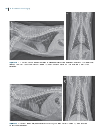
Figure 10.1 A 14-year-old domestic shorthair presented for lameness of left rear limb of one week duration. No heart murmur was
detected. The thoracic radiographic images are normal. The echocardiogram is normal. (a) Lateral projection. (b) Ventrodorsal
projection.
(b)
(a)
Figure 10.2 A 6-year-old Maine Coon presented for seizures. Radiographs of the thorax are normal. (a) Lateral projection.
(b) Ventrodorsal projection.