
Page 163 - Feline diagnostic imaging
P. 163
164 10 Normal Cardiovascular Imaging
Figure 10.4 Image showing hand position for scanning a
standing or sitting cat to obtain right and left ventricular short Figure 10.7 A right parasternal short axis view at the level of
axis views. the mitral valve leaflets typically called the “fish-mouth” view.
Right ventricle (RV) in the near field, with the left ventricle (LV)
and mitral valves (MV) depicted in the far field.
Figure 10.5 Image showing hand position for scanning a cat in
right lateral recumbency to obtain right and left ventricular
short axis views.
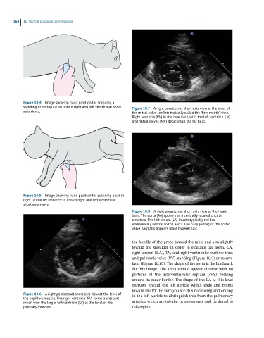
Figure 10.8 A right parasternal short axis view at the heart
base. The aorta (Ao) appears as a centrally located circular
structure. The left atrium (LA) in cats typically resides
immediately ventral to the aorta. The cusp (arroe) of the aortic
valve normally appears more hyperechoic.
the handle of the probe toward the table and aim slightly
toward the shoulder in order to evaluate the aorta, LA,
right atrium (RA), TV, and right ventricular outflow tract
and pulmonic valve (PV) standing (Figure 10.9) or recum-
bent (Figure 10.10). The shape of the aorta is the landmark
for this image. The aorta should appear circular with no
portions of the interventricular septum (IVS) peeking
around its outer border. The shape of the LA at this level
narrows toward the left auricle which ends and points
toward the PV. Be sure you see this narrowing and ending
Figure 10.6 A right parasternal short axis view at the level of in the left auricle to distinguish this from the pulmonary
the papillary muscle. The right ventricle (RV) forms a crescent
moon over the larger left ventricle (LV) at the level of the arteries, which are tubular in appearance and lie dorsal to
papillary muscles. this region.