
Page 48 - Feline diagnostic imaging
P. 48
42 4 Nuclear Imaging
While ultrasound and other cross‐sectional imaging should be normally hydrated before the examination and
modalities have largely replaced morphologic renal scintig- must remain motionless during the procedure so chemical
raphy, determination of GFR is likely the second most com- restraint is usually required. Anesthesia and sedation can
monly employed scintigraphic procedure in cats. Renal alter GFR, but studies have shown that acepromazine and
insufficiency and renal failure are common in older cats butorphanol can be used for restraint of cats without
and are a concern in cats treated for hyperthyroidism. While adversely affecting GFR results. Patients are positioned for
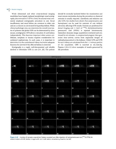
clinical pathologic data can determine the overall renal sta- dorsal imaging and 1–3 mCi of 99m Tc diethylenetriamine
tus of patients and global GFR can be determined by other pentaacetic acid (DTPA) is injected intravenously.
means, scintigraphic GFR allows evaluation of each kidney Immediate dynamic image acquisition is initiated and con-
independently. This becomes important when severe uro- tinued for six minutes. A computerized program then gen-
lithiasis, neoplasia or trauma requires consideration for erates time–activity curves from sequential images of
unilateral nephrectomy. In such cases, it is important to radiopharmaceutical in the kidneys. Global GFR and GFR
determine that the contralateral kidney maintains adequate for each kidney are calculated from the 1–3‐minute portion
function for survival if the affected kidney is removed. of the acquisition. GFR is reported as mL/min/kg.
Scintigraphy is a rapid, well‐documented and reliable Figures 4.10–4.12 show examples of results generated by
method to determine GFR in cats [19, 20]. The patient the procedure.
Figure 4.10 A series of dynamic acquisition frames acquired just after injection of radiopharmaceutical ( 99m Tc DTPA) for
determination of GFR. Ventral images with cat in left lateral recumbency, head to the left.