
Page 45 - Feline diagnostic imaging
P. 45
4.3 enal Scintigrappy 39
Several recent publications have evaluated 131 I dose
determination and effectiveness based on scoring systems
using combinations of the above criteria but none of these
has resulted in a method which ensures an appropriate
dose that will result in euthyroidism while voiding hypo-
thyroidism in all cats [12–14]. Some treatment centers use
a standard dose for all cats, some have a dose scale depend-
ing on the T4 level, and others use all information available
to individualize the dose for each cat.
Thyroid scintigraphy is by far the most commonly used
nuclear imaging procedure in cats [12, 15]. While scintigra-
phy is not essential for treatment of feline hyperthyroidism
and is not regularly used in many treatment centers, it does
offer useful information regarding the extent of disease. It
may be especially useful in confirming or excluding diag-
nosis in occult or questionable cases [16] and for evalua-
tion of treatment response when necessary. While thyroid
carcinoma cannot be definitively diagnosed by imaging,
scintigraphic patterns can suggest a higher likelihood of
malignancy and in those cases patterns associated with
malignancy include very large or multifocal sites of uptake,
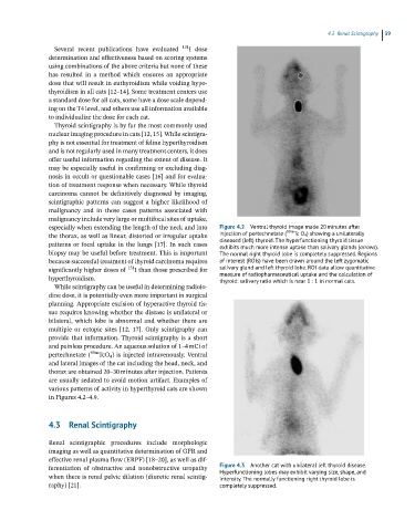
especially when extending the length of the neck and into Figure 4.2 Ventral thyroid image made 20 minutes after
the thorax, as well as linear, distorted or irregular uptake injection of pertechnetate ( 99m Tc O 4 ) showing a unilaterally
patterns or focal uptake in the lungs [17]. In such cases diseased (left) thyroid. The hyperfunctioning thyroid tissue
exhibits much more intense uptake than salivary glands (arrows).
biopsy may be useful before treatment. This is important The normal right thyroid lobe is completely suppressed. Regions
because successful treatment of thyroid carcinoma requires of interest (ROIs) have been drawn around the left zygomatic
significantly higher doses of 131 I than those prescribed for salivary gland and left thyroid lobe. ROI data allow quantitative
hyperthyroidism. measure of radiopharmaceutical uptake and the calculation of
thyroid: salivary ratio which is near 1 : 1 in normal cats.
While scintigraphy can be useful in determining radioio-
dine dose, it is potentially even more important in surgical
planning. Appropriate excision of hyperactive thyroid tis-
sue requires knowing whether the disease is unilateral or
bilateral, which lobe is abnormal and whether there are
multiple or ectopic sites [12, 17]. Only scintigraphy can
provide that information. Thyroid scintigraphy is a short
and painless procedure. An aqueous solution of 1–4 mCi of
pertechnetate ( 99m TcO 4 ) is injected intravenously. Ventral
and lateral images of the cat including the head, neck, and
thorax are obtained 20–30 minutes after injection. Patients
are usually sedated to avoid motion artifact. Examples of
various patterns of activity in hyperthyroid cats are shown
in Figures 4.2–4.9.
4.3 Renal Scintigraphy
Renal scintigraphic procedures include morphologic
imaging as well as quantitative determination of GFR and
effective renal plasma flow (ERPF) [18–20], as well as dif-
ferentiation of obstructive and nonobstructive uropathy Figure 4.3 Another cat with unilateral left thyroid disease.
Hyperfunctioning lobes may exhibit varying size, shape, and
when there is renal pelvic dilation (diuretic renal scintig- intensity. The normally functioning right thyroid lobe is
raphy) [21]. completely suppressed.