
Page 41 - Feline diagnostic imaging
P. 41
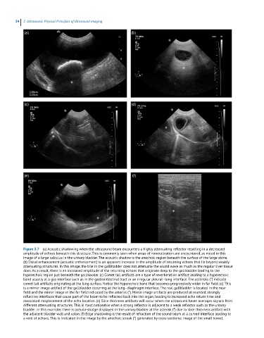
34  3  Ultrasound: Physical Principles of Ultrasound Imaging


            Figure 3.7  (a) Acoustic shadowing when the ultrasound beam encounters a highly attenuating reflector resulting in a decreased
            amplitude of echoes beneath this structure. This is commonly seen when areas of mineralization are encountered, as noted in this
            image of a large calculus in the urinary bladder. The acoustic shadow is the anechoic region beneath the surface of the large stone.
            (b) Distal enhancement (acoustic enhancement) is an apparent increase in the amplitude of returning echoes that lie beyond weakly
            attenuating structures. In this image, the bile in the gallbladder does not attenuate the sound wave as much as the regular liver tissue
            does. As a result, there is an increased amplitude of the returning echoes that originate deep to the gallbladder leading to the
            hyperechoic region just beneath the gallbladder. (c) Comet tail artifacts are a type of reverberation artifact leading to a hyperechoic
            band usually at a gas interface such as in the gastrointestinal tract or an irregular pleural–lung interface. The asterisks (*) indicate
            comet tail artifacts originating at the lung surface. Notice the hyperechoic band that becomes progressively wider in far field. (d) This
            is a mirror image artifact of the gallbladder occurring at the lung–diaphragm interface. The real gallbladder is located in the near
            field and the mirror image in the far field indicated by the asterisk (*). Mirror image artifacts are produced at rounded, strongly
            reflective interfaces that cause part of the beam to be reflected back into the organ, leading to increased echo return time and
            associated misplacement of the echo location. (e) Slice thickness artifacts will occur when the ultrasound beam averages signals from
            different attenuating structures. This is most noticeable when a strong reflector is adjacent to a weak reflector such as the urinary
            bladder. In this example, there is pseudo-sludge displayed in the urinary bladder at the asterisk (*) due to slice thickness artifact with
            the adjacent bladder wall and colon. (f) Edge shadowing is the result of refraction of the sound beam at a curved interface leading to
            a void of echoes. This is indicated in the image by the anechoic streak (*) generated by cross-sectional image of the small bowel.