
Page 37 - Feline diagnostic imaging
P. 37
30 3 Ultrasound: Physical Principles of Ultrasound Imaging
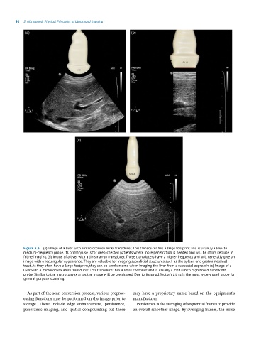
Figure 3.3 (a) Image of a liver with a macroconvex array transducer. This transducer has a large footprint and is usually a low- to
medium-frequency probe. Its primary use is for deep-chested patients where more penetration is needed and will be of limited use in
feline imaging. (b) Image of a liver with a linear array transducer. These transducers have a higher frequency and will generally give an
image with a rectangular appearance. They are valuable for imaging superficial structures such as the spleen and gastrointestinal
tract. As they often have a large footprint, they can be cumbersome when imaging the liver from a subcostal approach. (c) Image of a
liver with a microconvex array transducer. This transducer has a small footprint and is usually a medium to high broad bandwidth
probe. Similar to the macroconvex array, the image will be pie shaped. Due to its small footprint, this is the most widely used probe for
general purpose scanning.
As part of the scan conversion process, various preproc- may have a proprietary name based on the equipment’s
essing functions may be performed on the image prior to manufacturer.
storage. These include edge enhancement, persistence, Persistence is the averaging of sequential frames to provide
panoramic imaging, and spatial compounding but these an overall smoother image. By averaging frames, the noise