
Page 38 - Feline diagnostic imaging
P. 38
3.5 Image isplay 31
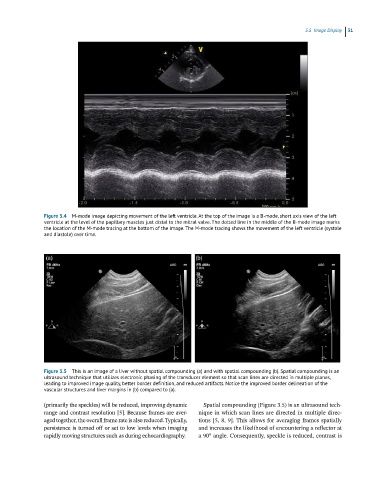
Figure 3.4 M-mode image depicting movement of the left ventricle. At the top of the image is a B-mode, short axis view of the left
ventricle at the level of the papillary muscles just distal to the mitral valve. The dotted line in the middle of the B-mode image marks
the location of the M-mode tracing at the bottom of the image. The M-mode tracing shows the movement of the left ventricle (systole
and diastole) over time.
Figure 3.5 This is an image of a liver without spatial compounding (a) and with spatial compounding (b). Spatial compounding is an
ultrasound technique that utilizes electronic phasing of the transducer element so that scan lines are directed in multiple planes,
leading to improved image quality, better border definition, and reduced artifacts. Notice the improved border delineation of the
vascular structures and liver margins in (b) compared to (a).
(primarily the speckles) will be reduced, improving dynamic Spatial compounding (Figure 3.5) is an ultrasound tech-
range and contrast resolution [5]. Because frames are aver- nique in which scan lines are directed in multiple direc-
aged together, the overall frame rate is also reduced. Typically, tions [5, 8, 9]. This allows for averaging frames spatially
persistence is turned off or set to low levels when imaging and increases the likelihood of encountering a reflector at
rapidly moving structures such as during echocardiography. a 90° angle. Consequently, speckle is reduced, contrast is