
Page 32 - Feline diagnostic imaging
P. 32
2.4 Magnetic Resonance Imaging 25
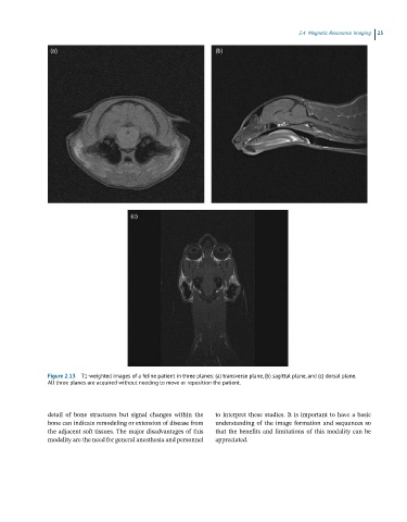
Figure 2.13 T1-weighted images of a feline patient in three planes: (a) transverse plane, (b) sagittal plane, and (c) dorsal plane.
All three planes are acquired without needing to move or reposition the patient.
detail of bone structures but signal changes within the to interpret these studies. It is important to have a basic
bone can indicate remodeling or extension of disease from understanding of the image formation and sequences so
the adjacent soft tissues. The major disadvantages of this that the benefits and limitations of this modality can be
modality are the need for general anesthesia and personnel appreciated.