
Page 31 - Feline diagnostic imaging
P. 31
24 2 Principles of Computed Tomography and Magnetic Resonance Imaging
Table 2.4 Different TR and TE sequences at a 1.5 T magnet depending on the area of interest. These sequences will
yield images that are T1, T2, or PD weighted. The entire
Sequence TE (ms) TR (ms) procedure can vary between 20 minutes to more than an
hour, depending on the amount of anatomy to be imaged.
T1 5–30 400–600 MR is an invaluable diagnostic tool particularly for the
T2 60–150 2000–4000 brain or spinal cord where more soft tissue detail is needed
PD 5–30 2000–4000 when evaluating for tumor, infections, inflammation, hem-
Source: Reference [1]. orrhage, or chronic disease. MR does not provide good
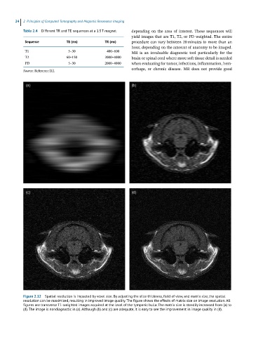
Figure 2.12 Spatial resolution is impacted by voxel size. By adjusting the slice thickness, field of view, and matrix size, the spatial
resolution can be maximized, resulting in improved image quality. The figure shows the effects of matrix size on image resolution. All
figures are transverse T1-weighted images acquired at the level of the tympanic bulla. The matrix size is steadily increased from (a) to
(d). The image is nondiagnostic in (a). Although (b) and (c) are adequate, it is easy to see the improvement in image quality in (d).