
Page 30 - Feline diagnostic imaging
P. 30
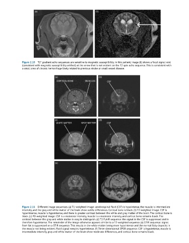
Figure 2.10  T2* gradient echo sequences are sensitive to magnetic susceptibility. In this patient, image (b) shows a focal signal void
               (consistent with magnetic susceptibility artifact) at the arrow that is not evident on the T2 spin echo sequence. This is consistent with
               a small area of chronic hemorrhage likely related to previous stroke or small vessel disease.


               Figure 2.11  Different image sequences. (a) T1-weighted image: cerebrospinal fluid (CSF) is hypointense, the muscle is intermediate
               intensity, and the gray and white matter of the brain show subtle differences. Cortical bone is black. (b) T2-weighted image: CSF is
               hyperintense, muscle is hypointense, and there is greater contrast between the white and gray matter of the brain. The cortical bone is
               black. (c) PD-weighted image: CSF is a moderate intensity, muscle is a moderate intensity, and cortical bone remains black. The
               contrast between the gray and white matter is easy to distinguish. (d) T2 FLAIR sequence: the signal in the CSF is suppressed and is
               therefore hypointense. The remainder of the image otherwise appears similar to a T2-weighted sequence. (e) STIR sequence: signal
               from fat is suppressed on a STIR sequence. This results in the white matter being more hypointense and the normal fatty deposits in
               the muscle not being evident. Fluid signal remains hyperintense. (f) Three-dimensional SPGR sequence: CSF is hypointense, muscle is
               intermediate intensity, gray and white matter of the brain show moderate differences, and cortical bone remains black.