
Page 25 - Feline diagnostic imaging
P. 25
18 2 Principles of Computed Tomography and Magnetic Resonance Imaging
pathology or to determine the extent of growth of a soft tis-
sue mass. Studies include but are not limited to the evalua-
tion of disease involving the nasal passages or tympanic
bullae, evaluation for metastasis or lung diseases, body
wall masses, head, or spinal trauma, retrobulbar and pitui-
tary masses. It is important to have a good understanding
of the CT principles as this technology is becoming more
mainstream.
2.4 Magnetic Resonance Imaging
2.4.1 MR Instrumentation
The MR system is a combination of magnets, coils, gradi-
ents, and computer control station. Through this combi-
nation, protons can be excited and spatially located, and
images generated. The main magnet is responsible for
generating the magnetic field. This magnet can be a high‐
field (>1 T) superconducting magnet or low‐field (<1 T)
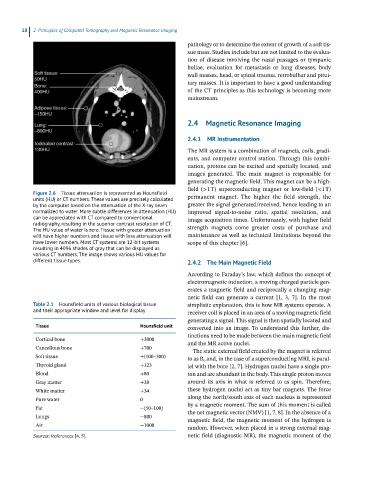
Figure 2.6 Tissue attenuation is represented as Hounsfield
units (HU) or CT numbers. These values are precisely calculated permanent magnet. The higher the field strength, the
by the computer based on the attenuation of the X-ray beam greater the signal generated/received, hence leading to an
normalized to water. More subtle differences in attenuation (HU) improved signal‐to‐noise ratio, spatial resolution, and
can be appreciated with CT compared to conventional image acquisition times. Unfortunately, with higher field
radiography, resulting in the superior contrast resolution of CT.
The HU value of water is zero. Tissue with greater attenuation strength magnets come greater costs of purchase and
will have higher numbers and tissue with less attenuation will maintenance as well as technical limitations beyond the
have lower numbers. Most CT systems are 12-bit systems scope of this chapter [6].
resulting in 4096 shades of gray that can be displayed as
various CT numbers. The image shows various HU values for
different tissue types. 2.4.2 The Main Magnetic Field
According to Faraday’s law, which defines the concept of
electromagnetic induction, a moving charged particle gen-
erates a magnetic field and reciprocally a changing mag-
netic field can generate a current [1, 3, 7]. In the most
Table 2.1 Hounsfield units of various biological tissue simplistic explanation, this is how MR systems operate. A
and their appropriate window and level for display receiver coil is placed in an area of a moving magnetic field
generating a signal. This signal is then spatially located and
Tissue Hounsfield unit converted into an image. To understand this further, dis-
tinctions need to be made between the main magnetic field
Cortical bone +3000 and the MR active nuclei.
Cancellous bone +700 The static external field created by the magnet is referred
Soft tissue +(100–300) to as B o and, in the case of a superconducting MRI, is paral-
Thyroid gland +123 lel with the bore [2, 7]. Hydrogen nuclei have a single pro-
Blood +80 ton and are abundant in the body. This single proton moves
Gray matter +38 around its axis in what is referred to as spin. Therefore,
White matter +34 these hydrogen nuclei act as tiny bar magnets. The force
Pure water 0 along the north/south axis of each nucleus is represented
Fat −(50–100) by a magnetic moment. The sum of this moment is called
Lungs −800 the net magnetic vector (NMV) [1, 7, 8]. In the absence of a
magnetic field, the magnetic moment of the hydrogen is
Air −1000 random. However, when placed in a strong external mag-
Sources: References [4, 5]. netic field (diagnostic MR), the magnetic moment of the