
Page 23 - Feline diagnostic imaging
P. 23
16 2 Principles of Computed Tomography and Magnetic Resonance Imaging
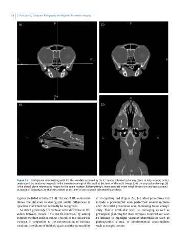
Figure 2.4 Multiplanar reformatting with CT. The raw data acquired by the CT can be reformatted in any plane to help viewers better
understand the anatomy. Image (b) is the transverse image of the skull at the level of the orbit. Image (c) is the sagittal and image (d)
is the dorsal plane reformatted image for the same location. Reformatting is more accurate when voxel dimensions are kept as small
as possible. Typically, slice thickness needs to be 1 mm or less to avoid reformatting artifacts.
regions are listed in Table 2.2 [4]. The use of HU values also of its capillary bed (Figure 2.8) [4]. Most procedures will
allows the clinician to distinguish subtle differences in include a postcontrast scan performed several minutes
opacities that would not normally be recognized. after the initial precontrast scan, increasing lesion conspi-
As stated previously, CT contrast is the difference in HU cuity. This is invaluable with neuroimaging as well as
values between tissues. This can be increased by adding presurgical planning for mass removal. Contrast can also
contrast medium such as iodine. The HU of the tissues will be utilized to highlight vascular abnormalities such as
increase in proportion to the concentration of contrast portosystemic shunts, or developmental abnormalities
medium, the volume of its blood space, and the permeability such as ectopic ureters.