
Page 145 - Veterinary Immunology, 10th Edition
P. 145
VetBooks.ir
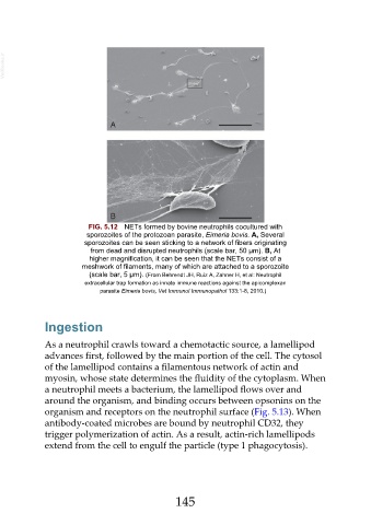
FIG. 5.12 NETs formed by bovine neutrophils cocultured with
sporozoites of the protozoan parasite, Eimeria bovis. A, Several
sporozoites can be seen sticking to a network of fibers originating
from dead and disrupted neutrophils (scale bar, 50 µm). B, At
higher magnification, it can be seen that the NETs consist of a
meshwork of filaments, many of which are attached to a sporozoite
(scale bar, 5 µm). (From Behrendt JH, Ruiz A, Zahner H, et al: Neutrophil
extracellular trap formation as innate immune reactions against the apicomplexan
parasite Eimeria bovis, Vet Immunol Immunopathol 133:1-8, 2010.)
Ingestion
As a neutrophil crawls toward a chemotactic source, a lamellipod
advances first, followed by the main portion of the cell. The cytosol
of the lamellipod contains a filamentous network of actin and
myosin, whose state determines the fluidity of the cytoplasm. When
a neutrophil meets a bacterium, the lamellipod flows over and
around the organism, and binding occurs between opsonins on the
organism and receptors on the neutrophil surface (Fig. 5.13). When
antibody-coated microbes are bound by neutrophil CD32, they
trigger polymerization of actin. As a result, actin-rich lamellipods
extend from the cell to engulf the particle (type 1 phagocytosis).
145