
Page 405 - Veterinary Immunology, 10th Edition
P. 405
VetBooks.ir
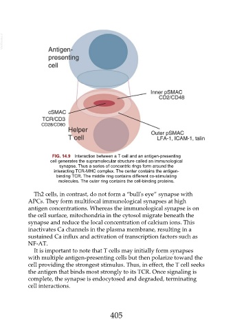
FIG. 14.9 Interaction between a T cell and an antigen-presenting
cell generates the supramolecular structure called an immunological
synapse. Thus a series of concentric rings form around the
interacting TCR-MHC complex. The center contains the antigen-
binding TCR. The middle ring contains different co-stimulating
molecules. The outer ring contains the cell-binding proteins.
Th2 cells, in contrast, do not form a “bull's eye” synapse with
APCs. They form multifocal immunological synapses at high
antigen concentrations. Whereas the immunological synapse is on
the cell surface, mitochondria in the cytosol migrate beneath the
synapse and reduce the local concentration of calcium ions. This
inactivates Ca channels in the plasma membrane, resulting in a
sustained Ca influx and activation of transcription factors such as
NF-AT.
It is important to note that T cells may initially form synapses
with multiple antigen-presenting cells but then polarize toward the
cell providing the strongest stimulus. Thus, in effect, the T cell seeks
the antigen that binds most strongly to its TCR. Once signaling is
complete, the synapse is endocytosed and degraded, terminating
cell interactions.
405