
Page 481 - Veterinary Immunology, 10th Edition
P. 481
VetBooks.ir
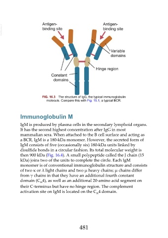
FIG. 16.3 The structure of IgG, the typical immunoglobulin
molecule. Compare this with Fig. 15.1, a typical BCR.
Immunoglobulin M
IgM is produced by plasma cells in the secondary lymphoid organs.
It has the second highest concentration after IgG in most
mammalian sera. When attached to the B cell surface and acting as
a BCR, IgM is a 180-kDa monomer. However, the secreted form of
IgM consists of five (occasionally six) 180-kDa units linked by
disulfide bonds in a circular fashion. Its total molecular weight is
then 900 kDa (Fig. 16.4). A small polypeptide called the J chain (15
kDa) joins two of the units to complete the circle. Each IgM
monomer is of conventional immunoglobulin structure and consists
of two κ or λ light chains and two µ heavy chains; µ chains differ
from γ chains in that they have an additional fourth constant
domain (C 4), as well as an additional 20-amino acid segment on
H
their C-terminus but have no hinge region. The complement
activation site on IgM is located on the C 4 domain.
H
481