
Page 484 - Veterinary Immunology, 10th Edition
P. 484
VetBooks.ir
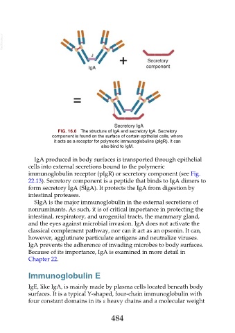
FIG. 16.6 The structure of IgA and secretory IgA. Secretory
component is found on the surface of certain epithelial cells, where
it acts as a receptor for polymeric immunoglobulins (pIgR). It can
also bind to IgM.
IgA produced in body surfaces is transported through epithelial
cells into external secretions bound to the polymeric
immunoglobulin receptor (pIgR) or secretory component (see Fig.
22.13). Secretory component is a peptide that binds to IgA dimers to
form secretory IgA (SIgA). It protects the IgA from digestion by
intestinal proteases.
SIgA is the major immunoglobulin in the external secretions of
nonruminants. As such, it is of critical importance in protecting the
intestinal, respiratory, and urogenital tracts, the mammary gland,
and the eyes against microbial invasion. IgA does not activate the
classical complement pathway, nor can it act as an opsonin. It can,
however, agglutinate particulate antigens and neutralize viruses.
IgA prevents the adherence of invading microbes to body surfaces.
Because of its importance, IgA is examined in more detail in
Chapter 22.
Immunoglobulin E
IgE, like IgA, is mainly made by plasma cells located beneath body
surfaces. It is a typical Y-shaped, four-chain immunoglobulin with
four constant domains in its ε heavy chains and a molecular weight
484