Page 148 - Manual of Equine Field Surgery
P. 148
144 HEAD AND NECK SURGERIES
site to minimize the tension on the tracheal
mucosa-skin junction during subsequent closure.
Bleeding is controlled with hemostat application
and or ligation if necessary. Subcutaneous tissue
may be sutured to the peritracheal fascia to
decrease dead space.
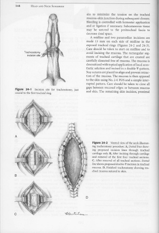
A midline and two paramedian incisions are
made 15 mm on each side of midline in the
exposed tracheal rings (Figures 24-2 and 24-3).
Care should be taken to start on midline and to
Tracheostomy avoid incising the mucosa. The rectangular seg-
incision site
ments of tracheal cartilage that are created are
carefully dissected free of mucosa. The mucosa is
desensitized with topical application of local anes-
thetic solution and incised in a double-Y-pattern.
Stay sutures are placed to align and prevent retrac-
tion of the mucosa. The mucosa is then apposed
to the skin using No. 2-0 PDS and a simple inter-
rupted pattern. Care should be taken to close all
gaps between mucosal edges or between mucosa
Figure 24-1 Incision site for tracheostomy, just
caudal to the first tracheal ring. and skin. The remaining skin incision, proximal
A
Figure 24-2 Ventral view of the neck illustrat-
ing tracheostomy procedure. A, Dotted lines show-
ing proposed incision lines through tracheal
cartilage only. B, After incising through cartilage
and removal of the first four tracheal sections.
C, After removal of all tracheal sections. Dotted
line shows proposed double- Y incision in tracheal
mucosa. D, Finished tracheostomy showing tra-
cheal mucosa sutured to skin.
B
D
c

