
Page 119 - Veterinary Histology of Domestic Mammals and Birds, 5th Edition
P. 119
Muscle tissue (textus muscularis) 101
VetBooks.ir
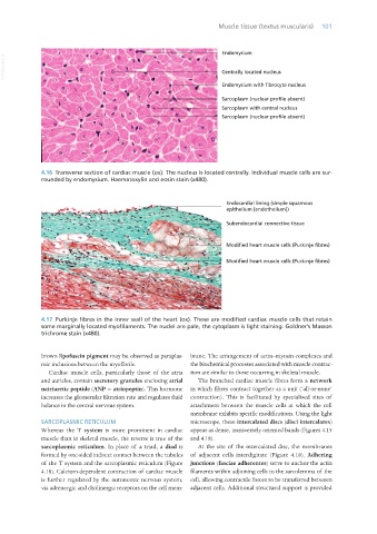
4.16 Transverse section of cardiac muscle (ox). The nucleus is located centrally. Individual muscle cells are sur-
rounded by endomysium. Haematoxylin and eosin stain (x480).
4.17 Purkinje fibres in the inner wall of the heart (ox). These are modified cardiac muscle cells that retain
some marginally located myofilaments. The nuclei are pale, the cytoplasm is light staining. Goldner’s Masson
trichrome stain (x480).
brown lipofuscin pigment may be observed as paraplas- brane. The arrangement of actin–myosin complexes and
mic inclusions between the myofibrils. the biochemical processes associated with muscle contrac-
Cardiac muscle cells, particularly those of the atria tion are similar to those occurring in skeletal muscle.
and auricles, contain secretory granules enclosing atrial The branched cardiac muscle fibres form a network
natriuretic peptide (ANP = atriopeptin). This hormone in which fibres contract together as a unit (‘all-or-none’
increases the glomerular filtration rate and regulates fluid contraction). This is facilitated by specialised sites of
balance in the central nervous system. attachment between the muscle cells at which the cell
membrane exhibits specific modifications. Using the light
SARCOPLASMIC RETICULUM microscope, these intercalated discs (disci intercalares)
Whereas the T system is more prominent in cardiac appear as dense, transversely oriented bands (Figures 4.15
muscle than in skeletal muscle, the reverse is true of the and 4.18).
sarcoplasmic reticulum. In place of a triad, a diad is At the site of the intercalated disc, the membranes
formed by one-sided indirect contact between the tubules of adjacent cells interdigitate (Figure 4.18). Adhering
of the T system and the sarcoplasmic reticulum (Figure junctions (fasciae adherentes) serve to anchor the actin
4.18). Calcium-dependent contraction of cardiac muscle filaments within adjoining cells to the sarcolemma of the
is further regulated by the autonomic nervous system, cell, allowing contractile forces to be transferred between
via adrenergic and cholinergic receptors on the cell mem- adjacent cells. Additional structural support is provided
Vet Histology.indb 101 16/07/2019 14:57