
Page 120 - Veterinary Histology of Domestic Mammals and Birds, 5th Edition
P. 120
102 Veterinary Histology of Domestic Mammals and Birds
VetBooks.ir
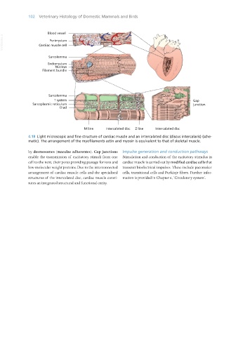
4.18 Light microscopic and fine structure of cardiac muscle and an intercalated disc (discus intercalaris) (sche-
matic). The arrangement of the myofilaments actin and myosin is equivalent to that of skeletal muscle.
by desmosomes (maculae adherentes). Gap junctions Impulse generation and conduction pathways
enable the transmission of excitatory stimuli from one Stimulation and conduction of the excitatory stimulus in
cell to the next, their pores providing passage for ions and cardiac muscle is carried out by modified cardiac cells that
low-molecular weight proteins. Due to the interconnected transmit bioelectrical impulses. These include pacemaker
arrangement of cardiac muscle cells and the specialised cells, transitional cells and Purkinje fibres. Further infor-
structures of the intercalated disc, cardiac muscle consti- mation is provided in Chapter 6, ‘Circulatory system’.
tutes an integrated structural and functional entity.
Vet Histology.indb 102 16/07/2019 14:57