
Page 136 - Veterinary Histology of Domestic Mammals and Birds, 5th Edition
P. 136
118 Veterinary Histology of Domestic Mammals and Birds
VetBooks.ir
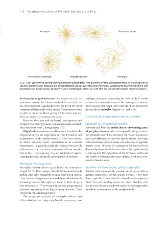
5.25 Glial cells of the central nervous system (schematic). The portions of the cell represented in the diagram as
continuous lines can typically be demonstrated using silver staining methods. Subsequent branching of the cell
processes can occasionally be shown with histological stains or with the aid of ultrastructural reconstructions.
Perivascular oligodendrocytes are numerous and are rophages, pericytes surrounding the walls of blood vessels
prominent around the blood vessels of the cerebral cor- or from the connective tissue of the meninges. In view of
tex. Interfascicular oligodendrocytes are by far the most their mesodermal origin, they have also been referred to
common cell type in the white matter. Their processes run historically as mesoglia (Figures 5.24 and 5.25).
parallel to the nerve fibres, giving off branches that par-
tially or completely surround the axon. Glial cells of the peripheral nervous system
Based on their size and the length, arrangement and
complexity of their processes, oligodendrocytes are subdi- Schwann cell (neurolemmocyte)
vided into four types (Hortega types I–IV). Schwann cells form the myelin sheath surrounding axons
Oligodendrocytes have several functions. Interfascicular of peripheral nerves. After coming to lie along an axon,
oligodendrocytes are responsible for the formation and the plasmalemma of the Schwann cell wraps around the
maintenance of the myelin sheath of CNS nerve fibres, axon and differentiates into the myelin sheath. Schwann
in which saltatory nerve conduction is an essential cells surround peripheral axons over a distance of approxi-
requirement. Oligodendrocytes also interact functionally mately 1 mm. The ends of consecutive Schwann cells are
with neurons and are a key component of iron metabo- denoted by the node of Ranvier, where the myelin sheath
lism in the CNS, requiring iron for synthesis of myelin. is interrupted. The cytoplasm of the Schwann cell forms
Oligodendrocytes inhibit the development of neurites. the sheath of Schwann, the outer surface of which is com-
prised of basal lamina.
Microglia (Hortega cells)
Microglia, also named Hortega cells after the neurophysi- Satellite cell (amphicyte, gliocytus ganglii)
ologist Pio Del Rio Hortega (1882–1945), are small, usually Satellite cells surround the perikaryon of nerve cells in
stellate glial cells, frequently located near blood vessels. ganglia (autonomic, spinal, cranial nerves). They have
They have an elongated nucleus and short, often bizarrely dense, typically flattened nuclei. A basal lamina separates
formed, processes. Microglia are found in both the grey them from surrounding connective tissue. Satellite cells
and white matter. They frequently contain phagocytosed are functionally associated with capillaries and support the
material originating from degenerating neurons. Their metabolic requirements of the ganglion cells.
cytoplasm contains phagosomes.
The phagocytic capacity of microglia reflects their
differentiation from migrating blood monocytes, mac-
Vet Histology.indb 118 16/07/2019 14:57