
Page 171 - Veterinary Histology of Domestic Mammals and Birds, 5th Edition
P. 171
Immune system and lymphatic organs (organa Iymphopoetica) 153
Based on structural and functional criteria, lymphoid Tonsils and Peyer’s patches
VetBooks.ir follicles are categorised as either primary or secondary. Tonsils are aggregations of individual lymphoid follicles,
Primary follicles contain an accumulation of mature, found particularly in the oropharyngeal region.
Tonsils are characterised by the following features:
yet incompletely differentiated (naïve), B lymphocytes
that have not yet come into contact with an antigen. Upon
antigenic stimulation, facilitated by reticular and dendritic · They are a component of the mucosa-associated
cells, these become immunologically active B cells. lymphatic tissue and contain primary and second-
Secondary follicles develop following activation of ary follicles.
primary follicles by contact with an antigen. They are · They are covered by epithelium; leucocytes are typi-
characterised by the presence of a germinal centre where cally present between epithelial cells.
proliferation and differentiation of B cells takes place. · Connective tissue surrounds (capsule) and subdi-
Follicular dendritic cells, macrophages and helper T cells vides the lymphoid tissue.
are also present in germinal centres; helper T cells play an · They are located near primarily mucous (seromu-
important role in B-cell differentiation. cous in carnivores) glands.
The germinal centre is composed of a light zone and · They have only efferent lymphatic vessels; afferent
an adjacent dark zone. A so-called mantle layer (corona) vessels are absent.
forms a thin layer around the light zone. After exposure
to an antigen, B lymphocytes proliferate in the dark zone Tonsils provide immune defence against antigens within
(usually located at one pole of the follicle). These prolifer- the oral and pharyngeal cavities. Based on location, they
ating B cells are referred to as centroblasts. Centroblasts are referred to as palatine tonsils, tonsils of the soft palate,
differentiate into centrocytes, which give the germinal pharyngeal tonsils, lingual tonsils, para-epiglottic tonsils or
centre its relatively light appearance. Centrocytes present tubal (Eustachian) tonsils. Structurally, tonsils have either
antigens on their surface. Those with a high affinity for a smooth or invaginated surface.
antigen subsequently develop into plasma cells or memory Those with a smooth surface, the simplest form of ton-
cells. The remainder are eliminated by apoptosis. sil, include the palatine tonsil of carnivores (Figure 8.5).
Secondary follicles thus serve as centres of prolifera- Other tonsils have deep fossules in their surface (fossula
tion of B cells and their active progeny, plasma cells. tonsillaris). These include the lingual tonsils of the horse,
In addition to occurring as solitary structures, lymphoid the tonsil of the soft palate of the horse and pig and the
follicles also manifest in aggregated forms – for example in tubal tonsils of the pig. The invaginations of the palatine
lymph nodes, the white pulp of the spleen, the tonsils and tonsil of ruminants are compound in nature, with the fos-
Peyer’s patches. sula opening into a branching tonsillar sinus (Figure 8.6).
Of particular note are the tonsil-like lymphatic organs of
the gut, the Peyer’s patches. These are found particularly
in the ileum, but also in the duodenum and jejunum, and in
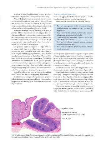
8.5 Palatine tonsil (dog). Tonsils are subepithelial accumulations of lymphoid follicles, enclosed in a capsule and
located near glandular tissue. Haematoxylin and eosin stain (x8).
Vet Histology.indb 153 16/07/2019 14:59