
Page 174 - Veterinary Histology of Domestic Mammals and Birds, 5th Edition
P. 174
156 Veterinary Histology of Domestic Mammals and Birds
meshwork of reticular fibres and reticular cells. The Vessels and sinuses
VetBooks.ir spaces within the meshwork are filled with cells including The function of lymph nodes is closely related to the
lymphocytes, plasma cells and macrophages (Figure 8.10).
structure of the lymph vessels and their distribution in
Based on the differential distribution of these cells, the cortex and medulla. After passing through the capsule,
lymph nodes are divided into regions designated as: the afferent lymph vessels empty into an expanded sub-
capsular sinus. This gives rise to sinuses (oriented towards
· an outer subcapsular cortex, the centre of the node) that accompany connective tissue
· the paracortex (thymus-dependent region) and trabeculae through the cortex (cortical or intermediate
· the inner medulla, composed of lymphoid cells and sinuses). The cortical sinuses continue into an extensive
reticular tissue (Figures 8.7 and 8.8). interconnected network of chambers, the medullary
sinuses. These converge to form efferent lymph vessels
The cortex consists largely of aggregations of primary and that leave the node at the hilus (Figure 8.7).
secondary follicles containing inactive and activated B lym- The walls of the sinuses are largely discontinu-
phocytes (Figures 8.7 to 8.11). Lying between and deep ous, facilitating the passage of lymphoid cells into the
to the lymphoid follicles is the paracortex (parafollicular parenchyma. The endothelial lining consists of modified
zone), in which T cells are concentrated. reticular cells. Macrophages (part of the MPS) are present
Within the paracortex, the evenly distributed T cells in the lumina of the sinuses. Antigens present in lymph can
are surrounded by interdigitating dendritic cells and likewise traverse the sinus walls to reach the parenchyma,
high endothelial venules. Introduction of an antigen where they interact with antigen presenting cells, leading
to a lymph node initiates T- and B-cell cooperation and to T-cell and macrophage activation.
maturation. Activated B cells (along with some T cells) Activated T lymphocytes convey information to other
migrate to the follicles where they undergo further matu- lymphocytes within the follicles, triggering a cascade of
ration before passing to the medulla as antigen-secreting cellular responses. In this process, the thymus-dependent
cells. paracortex becomes enlarged and the germinal centres
The medulla contains connective tissue trabeculae develop through the activation of B cells and formation of
through which numerous small blood vessels pass. The plasma cells and memory cells. The lymph node increases
parenchyma consists of medullary cords (clusters of cells in size. Activated plasma cells migrate into the medullary
including lymphocytes, plasma cells and macrophages) sinuses where they release specific antibodies into the lymph.
separated by medullary lymph sinuses (see below). The
cords are reinforced by a reticular meshwork. Species variation
Lymph nodes are involved in active filtration of lymph Pig: The structure of the lymph nodes of the pig differs
before it returns to the circulating blood. Cells of the immune from that of other mammals (Figure 8.9), the main dis-
system eliminate foreign and damaged endogenous cells via tinction being the arrangement of the lymph vessels. In
innate and adaptive mechanisms. Mononuclear phagocytes porcine lymph nodes, the afferent lymph vessels enter at
(macrophages) engulf cell debris, bacteria, viruses, toxins the hilus and the efferent vessels leave the node through
and exogenous pigments. T and B cells are responsible for the capsule.
cytotoxicity and specific antibody production.
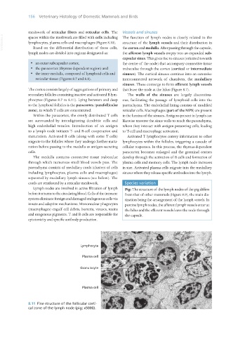
8.11 Fine structure of the follicular corti-
cal zone of the lymph node (pig; x5000).
Vet Histology.indb 156 16/07/2019 14:59